
Stomatology ›› 2025, Vol. 45 ›› Issue (11): 839-843.doi: 10.13591/j.cnki.kqyx.2025.11.007
• Basic and Clinical Research • Previous Articles Next Articles
LIU Yi1, ZHOU Xiang1, SHEN Dong1, CHU Manru2( ), WEI Changbo3
Received:2025-01-02
Online:2025-11-28
Published:2025-11-18
Contact:
CHU Manru
E-mail:chumanru2010@126.com
CLC Number:
LIU Yi, ZHOU Xiang, SHEN Dong, CHU Manru, WEI Changbo. CBCT study on the anatomical structure related to immediate implantation of maxillary premolars[J]. Stomatology, 2025, 45(11): 839-843.
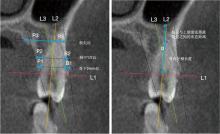
Fig.1
Measurements of maxillary premolars"

Tab.1
The thickness of buccal and palatal bone plate in maxillary premolar mm"
| 牙位 | 测量位置 | 颊侧骨壁 | 腭侧骨壁 | Z | P |
|---|---|---|---|---|---|
| 上颌第一前磨牙 | 骨下2 mm处 | 1.36(0.88,1.84) | 1.20(0.92,1.65) | -1.295 | 0.195 |
| 根中1/2处 | 0.96(0.73,1.65) | 1.97(1.61,2.64) | -5.995 | <0.001 | |
| 根尖处 | 0.73(0.23,1.50) | 7.35(5.68,9.33) | -7.913 | <0.001 | |
| 上颌第二前磨牙 | 骨下2 mm处 | 1.84(1.32,2.37) | 1.25(0.98,1.56) | -4.311 | <0.001 |
| 根中1/2处 | 1.96(1.30,2.53) | 2.16(1.61,2.56) | -1.666 | 0.096 | |
| 根尖处 | 2.83(1.34,4.18) | 6.87(5.57,8.23) | -6.896 | <0.001 |
Tab.2
Comparison of the thickness of buccal and palatal bone plate in different genders mm"
| 牙位 | 测量位置 | 性别 | t/Z | P | |
|---|---|---|---|---|---|
| 男 | 女 | ||||
| 上颌第一前磨牙 | B1 | 1.48±0.85 | 1.35±0.61 | 0.790 | 0.430 |
| B2 | 1.24(0.72,2.04) | 0.95(0.74,1.43) | -1.301 | 0.193 | |
| B3 | 0.92(0.62,1.65) | 0.45(0.00,1.29) | -2.656 | <0.010 | |
| P1 | 1.39±0.63 | 1.16±0.45 | -1.419 | 0.156 | |
| P2 | 2.20±0.83 | 2.01±0.81 | 1.040 | 0.300 | |
| P3 | 7.98(6.09,10.16) | 6.85(5.44,8.29) | -1.729 | 0.084 | |
| 上颌第二前磨牙 | B1 | 1.94±0.78 | 1.84±0.72 | 0.590 | 0.560 |
| B2 | 2.08±0.92 | 1.91±0.87 | 0.850 | 0.400 | |
| B3 | 3.27(2.46,4.29) | 2.30(0.90,4.05) | -2.190 | 0.029 | |
| P1 | 1.25(1.05,1.87) | 1.24(0.94,1.48) | -0.680 | 0.497 | |
| P2 | 2.39(1.84,2.98) | 1.74(1.44,2.40) | -3.057 | <0.010 | |
| P3 | 7.57(6.46,8.57) | 6.18(4.57,7.60) | -2.190 | 0.040 | |
Tab.3
Correlation analysis between the angle α and the thickness of the buccal and palatal bone plate of the maxillary premolar"
| 测量项目 | 相关系数 | P |
|---|---|---|
| B1 | -0.036 | 0.631 |
| B2 | -0.185 | 0.014 |
| B3 | -0.516 | <0.001 |
| P1 | -0.040 | 0.595 |
| P2 | 0.201 | 0.008 |
| P3 | 0.460 | <0.001 |
| [1] |
Schulte W, Heimke G. The tübinger immediate implant[J]. Quintessenz, 1976, 27(6): 17-23.
pmid: 802007 |
| [2] |
Fan R, Quinton HA, Golberg MB, et al. Immediate implants[J]. Dent Clin N Am, 2021, 65(1): 89-102.
doi: 10.1016/j.cden.2020.09.007 |
| [3] |
Mark I, Dym H, Fan YJ. Immediate restoration of an endosseous implant[J]. Dent Clin North Am, 2024, 68(1): 203-212.
doi: 10.1016/j.cden.2023.08.002 pmid: 37951633 |
| [4] |
Ahmad R. Is there an advantage to delayed molar implant placement in those with chronic apical periodontitis?[J]. Evid Based Dent, 2024, 25(2): 79-80.
doi: 10.1038/s41432-024-00993-w pmid: 38531998 |
| [5] |
Rojas-Vizcaya F. Biological aspects as a rule for single implant placement. The 3A-2B rule: A clinical report[J]. J Prosthodont, 2013, 22(7): 575-580.
doi: 10.1111/jopr.12039 pmid: 23551872 |
| [6] |
Liñares A, Dopico J, Magrin G, et al. Critical review on bone grafting during immediate implant placement[J]. Periodontol 2000, 2023, 93(1): 309-326.
doi: 10.1111/prd.12516 pmid: 37658586 |
| [7] | 童丽, 顾卫平, 陈岗, 等. 基于CBCT的下颌第一磨牙区即刻种植相关的影像学研究[J]. 口腔医学, 2020, 40(3): 227-231. |
| [8] |
丁子凌, 刘昕, 杨晓喻, 等. 华南地区成人上颌中切牙与牙槽骨相对位置关系CBCT分析[J]. 口腔疾病防治, 2024, 32 (2): 116-122.
doi: 10.12016/j.issn.2096-1456.2024.02.005 |
| [9] |
Saleh Hasani Jebelli M, Yari A, Nikparto N, et al. Tissue engineering innovations to enhance osseointegration in immediate dental implant loading: A narrative review[J]. Cell Biochem Funct, 2024, 42(2):e3974.
doi: 10.1002/cbf.3974 pmid: 38491807 |
| [10] | Vasiljevic M, Selakovic D, Rosic G, et al. Anatomical factors of the anterior and posterior maxilla affecting immediate implant placement based on cone beam computed tomography analysis: A narrative review[J]. Diagnostics (Basel), 2024, 14(15): 1697. |
| [11] | Patel R, Ucer C, Wright S, et al. Differences in dental implant survival between immediate vs. delayed placement: A systematic review and meta-analysis[J]. Dent J (Basel), 2023, 11(9): 218. |
| [12] |
Zhao G, Zhou Y, Shi S, et al. Long-term clinical outcomes of immediate loading versus non-immediate loading in single-implant restorations: A systematic review and meta-analysis[J]. Int J Oral Maxillofac Surg, 2022, 51(10): 1345-1354.
doi: 10.1016/j.ijom.2022.03.057 |
| [13] | Alqutaibi AY, Aloufi AM, Hamadallah HH, et al. Multifactorial analysis of the maxillary premolar area for immediate implant placement using cone beam computed tomography: A study of 333 maxillary images[J]. J Prosthet Dent, 2024: S0022-3913(24)00468-2. |
| [14] |
Stoilov M, Shafaghi R, Stark H, et al. Influence of implant macro-design, -length, and-diameter on primary implant stability depending on different bone qualities using standard drilling protocols-an in vitro analysis[J]. J Funct Biomater, 2023, 14(9): 469.
doi: 10.3390/jfb14090469 |
| [15] |
von Arx T, Fodich I, Bornstein MM. Proximity of premolar roots to maxillary sinus: A radiographic survey using cone-beam computed tomography[J]. J Endod, 2014, 40(10): 1541-1548.
doi: 10.1016/j.joen.2014.06.022 pmid: 25129024 |
| [16] |
刘昕, 丁子凌, 杨晓喻, 等. 锥形束CT测量成人上颌前磨牙根尖与上颌窦位置关系及其对即刻种植的影响[J]. 口腔疾病防治, 2024, 32(6): 444-450.
doi: 10.12016/j.issn.2096-1456.2024.06.006 |
| [17] | 韦梦瑶, 王晓丽, 李雁, 等. 上颌前磨牙根管变异及其与上颌窦关系的锥形束CT研究[J]. 上海口腔医学, 2018, 27(2): 156-163. |
| [18] |
Huang YC, Huang YC, Ding SJ. Primary stability of implant placement and loading related to dental implant materials and designs: A literature review[J]. J Dent Sci, 2023, 18(4): 1467-1476.
doi: 10.1016/j.jds.2023.06.010 |
| [19] |
Najm A, Bihorac A, de Carvalho Machado V, et al. Immediate implant placement in the premolar maxillary area: A cone-beam computed tomography study[J]. J Periodontal Implant Sci, 2025, 55(1): 72-84.
doi: 10.5051/jpis.2303580179 |
| [20] | 王驰, 任秀云. 唇侧骨壁缺损情况下即刻种植的研究进展[J]. 口腔医学, 2023, 43(12): 1140-1144. |
| [21] | 宋颢, 杨梦源, 陆艺文, 等. 上前牙区种植相关解剖因素CBCT研究[J]. 中国临床解剖学杂志, 2023, 41(4): 401-408. |
| [22] |
Pitman J, Christiaens V, Callens J, et al. Immediate implant placement with flap or flapless surgery: A systematic review and meta-analysis[J]. J Clin Periodontol, 2023, 50(6): 755-764.
doi: 10.1111/jcpe.v50.6 |
| [23] |
Yan YJ, Li JL, Zhu HL, et al. CBCT evaluation of root canal morphology and anatomical relationship of root of maxillary second premolar to maxillary sinus in a western Chinese population[J]. BMC Oral Health, 2021, 21(1): 358.
doi: 10.1186/s12903-021-01714-w pmid: 34284763 |
| Viewed | ||||||
|
Full text |
|
|||||
|
Abstract |
|
|||||