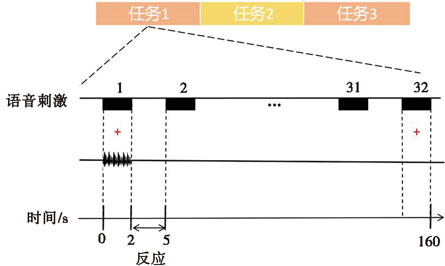
| [1] |
Paal S, Reulbach U, Strobel-Schwarthoff K, et al. Evaluation of speech disorders in children with cleft lip and palate[J]. J Orofac Orthop, 2005, 66(4):270-278.
pmid: 16044225
|
| [2] |
李杨, 尹恒. 腭裂语音与评估[M]. 北京: 人民军医出版社, 2015.
|
| [3] |
Peterson-Falzone S, Trost-Cardamone J, Karnell M, et al. The clinician's guide to treating cleft palate speech[M]. 2nd Edition. Amsterdam: Elsvier, 2016.
|
| [4] |
韩源, 汪彬昺, 李盛, 等. 一种以治疗方式为导向的腭裂相关语音障碍的分型系统:基于347例患者的回顾研究[J]. 口腔医学, 2022, 42(4):332-338.
|
| [5] |
Nopoulos P, Berg S, Canady J, et al. Structural brain abnormalities in adult males with clefts of the lip and/or palate[J]. Genet Med, 2002, 4(1):1-9.
doi: 10.1097/00125817-200201000-00001
pmid: 11839951
|
| [6] |
Nopoulos P, Choe I, Berg S, et al. Ventral frontal cortex morphology in adult males with isolated orofacial clefts: Relationship to abnormalities in social function[J]. Cleft Palate Craniofac J, 2005, 42(2):138-144.
doi: 10.1597/03-112.1
|
| [7] |
Nopoulos P, Berg S, VanDemark D, et al. Increased incidence of a midline brain anomaly in patients with nonsyndromic clefts of the lip and/or palate[J]. J Neuroimaging, 2001, 11(4):418-424.
pmid: 11677883
|
| [8] |
Adamson CL, Anderson VA, Nopoulos P, et al. Regional brain morphometric characteristics of nonsyndromic cleft lip and palate[J]. Dev Neurosci, 2014, 36(6):490-498.
doi: 10.1159/000365389
|
| [9] |
Boes AD, Murko V, Wood JL, et al. Social function in boys with cleft lip and palate: Relationship to ventral frontal cortex morphology[J]. Behav Brain Res, 2007, 181(2):224-231.
pmid: 17537526
|
| [10] |
Zhang WJ, Zhao C, Sun LW, et al. Articulation-function-associated cortical developmental changes in patients with cleft lip and palate[J]. Brain Sci, 2023, 13(4):550.
doi: 10.3390/brainsci13040550
|
| [11] |
Becker DB, Coalson RS, Sachanandani NS, et al. Functional neuroanatomy of lexical processing in children with cleft lip and palate[J]. Plast Reconstr Surg, 2008, 122(5):1371-1382.
doi: 10.1097/PRS.0b013e3181881f54
pmid: 18971720
|
| [12] |
Goldsberry G, O'Leary D, Hichwa R, et al. Functional abnormalities in the neural circuitry of reading in men with nonsyndromic clefts of the lip or palate[J]. Cleft Palate Craniofac J, 2006, 43(6):683-690.
doi: 10.1597/05-043
|
| [13] |
张文婧, 赵翠, 李春林, 等. 腭裂言语障碍儿童语言相关脑区灰质形态学分析[J]. 中华口腔医学杂志, 2022, 57(9):899-906.
|
| [14] |
Li Z, Zhang WJ, Li CL, et al. Articulation rehabilitation induces cortical plasticity in adults with non-syndromic cleft lip and palate[J]. Aging, 2020, 12(13):13147-13159.
doi: 10.18632/aging.v12i13
|
| [15] |
Rao B, Cheng H, Xu HB, et al. Random network and non-rich-club organization tendency in children with non-syndromic cleft lip and palate after articulation rehabilitation: A diffusion study[J]. Front Neurol, 2022, 13: 790607.
doi: 10.3389/fneur.2022.790607
|
| [16] |
Zhang WJ, Li CL, Chen L, et al. Increased activation of the hippocampus during a Chinese character subvocalization task in adults with cleft lip and palate palatoplasty and speech therapy[J]. Neuroreport, 2017, 28(12):739-744.
doi: 10.1097/WNR.0000000000000832
pmid: 28658048
|
| [17] |
Peck KK, Galgano JF, Branski RC, et al. Event-related functional MRI investigation of vocal pitch variation[J]. Neuro Image, 2009, 44(1):175-181.
|
| [18] |
Euston DR, Gruber AJ, McNaughton BL. The role of medial prefrontal cortex in memory and decision making[J]. Neuron, 2012, 76(6):1057-1070.
doi: 10.1016/j.neuron.2012.12.002
pmid: 23259943
|
| [19] |
Wei JW, Zhang ZG, Yao ZQ, et al. Modulation of sustained attention by Theta-tACS over the lateral and medial frontal cortices[J]. Neural Plast, 2021, 2021: 5573471.
|
| [20] |
Bai Y, Liu S, Zhu M, et al. Perceptual pattern of cleft-related speech: A task-fMRI study on typical mandarin-speaking adults[J]. Brain Sci, 2023, 13(11):1506.
doi: 10.3390/brainsci13111506
|
| [21] |
McGugin RW, Ryan KF, Tamber-Rosenau BJ, et al. The role of experience in the face-selective response in right FFA[J]. CerebCortex, 2018, 28(6):2071-2084.
|
| [22] |
van Atteveldt N, Murray MM, Thut G, et al. Multisensory integration: Flexible use of general operations[J]. Neuron, 2014, 81(6):1240-1253.
doi: S0896-6273(14)00194-9
pmid: 24656248
|
| [23] |
Opoku-Baah C, Schoenhaut AM, Vassall SG, et al. Visual influences on auditory behavioral, neural, and perceptual processes: A review[J]. JAssocResOtolaryngol, 2021, 22(4):365-386.
|
| [24] |
Breedlove JL, St-Yves G, Olman CA, et al. Generative feedback explains distinct brain activity codes for seen and mental images[J]. Curr Biol, 2020, 30(12):2211-2224.e6.
doi: S0960-9822(20)30494-2
pmid: 32359428
|
| [25] |
Arsenault JS, Buchsbaum BR. No evidence of somatotopic place of articulation feature mapping in motor cortex during passive speech perception[J]. Psychon Bull Rev, 2016, 23(4):1231-1240.
doi: 10.3758/s13423-015-0988-z
pmid: 26715582
|
| [26] |
Vetter P, Smith FW, Muckli L. Decoding sound and imagery content in early visual cortex[J]. Curr Biol, 2014, 24(11):1256-1262.
doi: 10.1016/j.cub.2014.04.020
pmid: 24856208
|
| [27] |
Jiang CH, Whitehill TL, McPherson B, et al. Spectral moment analysis of affricates produced by Mandarin-speaking pre-adolescents with repaired cleft palate[J]. Int J Pediatr Otorhinolaryngol, 2016, 84: 137-142.
doi: 10.1016/j.ijporl.2016.01.029
|
| [28] |
Güntürkün O, Ströckens F, Ocklenburg S. Brain lateralization: A comparative perspective[J]. Physiol Rev, 2020, 100(3):1019-1063.
doi: 10.1152/physrev.00006.2019
pmid: 32233912
|
| [29] |
王帅. 语言功能偏侧化的脑网络研究[D]. 上海: 华东师范大学, 2018.
|
| [30] |
Schneider HR, Wawrzyniak M, Stockert A, et al. fMRI informed voxel-based lesion analysis to identify lesions associated with right-hemispheric activation in aphasia recovery[J]. Neuroimage Clin, 2022, 36: 103169.
doi: 10.1016/j.nicl.2022.103169
|
| [31] |
Deng XF, Wang B, Zong FR, et al. Right-hemispheric language reorganization in patients with brain arteriovenous malformations: A functional magnetic resonance imaging study[J]. Hum Brain Mapp, 2021, 42(18):6014-6027.
doi: 10.1002/hbm.25666
pmid: 34582074
|

 ),江宏兵1(