关闭×
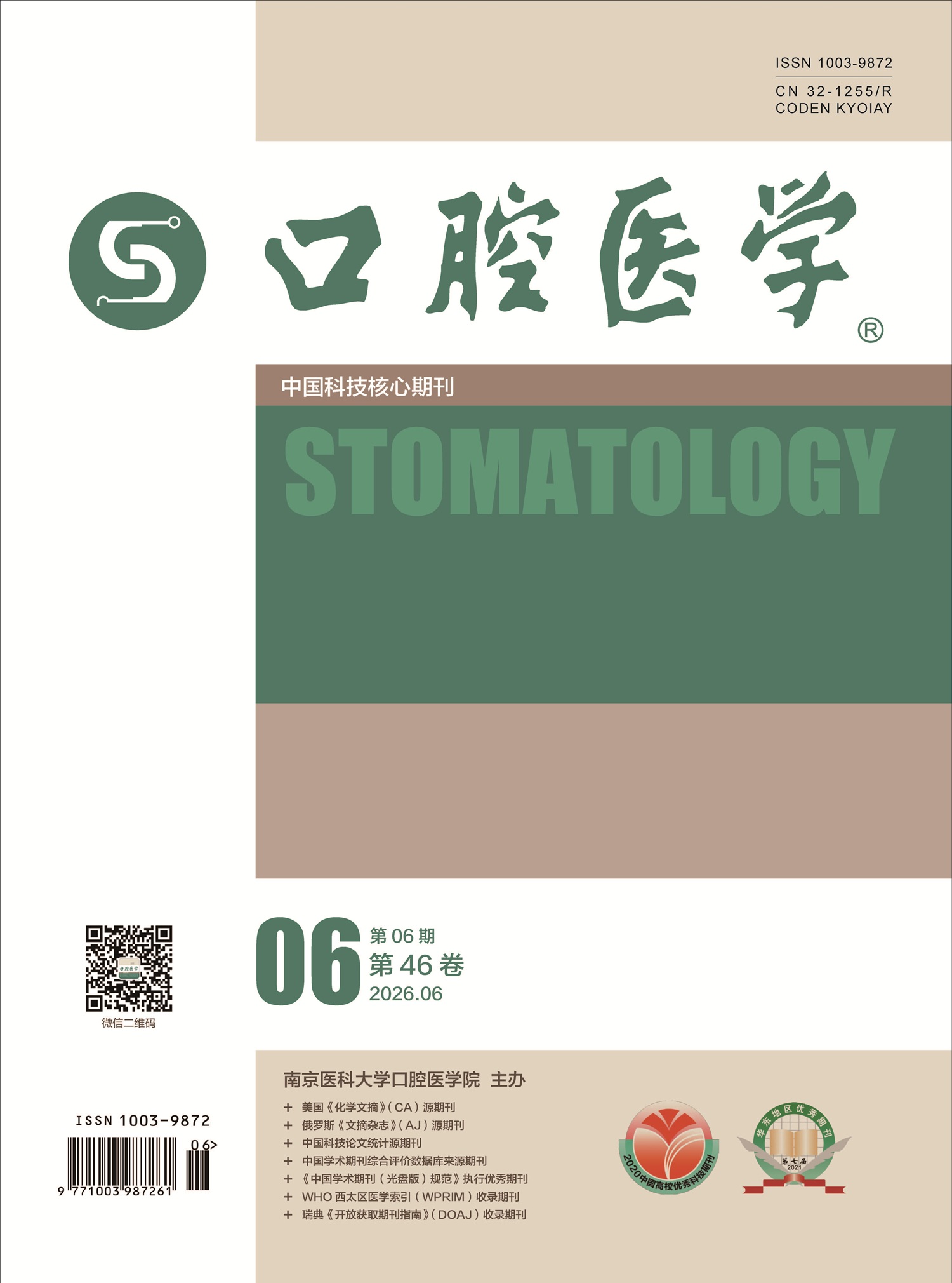
 Stomatology, established in 1981, has a history of more than 40 years and is the second comprehensive journal of stomatology in China. The journal is sponsored by Stomatological College of Nanjing Medical University, and co-sponsored by the School of Stomatology of Shandong University, School of Stomatology of Tongji University, Changzhou Stomatological Hospital of Jiangsu Province, Stomatology Hospital, Zhejiang University School of Medicine, and Zhejiang Stomatological Association.
In 2003, the journal was included in “Source Journals of Chinese Scientific and Technological ...更多
Current Issue
28 January 2025, Volume 45 Issue 1
Members of the Chinese Academy of Medical Sciences Column
Maternal oral-gut microbiome and artificial assisted reproduction
YANG Peiyue, PENG Xian, ZHOU Xuedong
2025, 45(1):  1-7.  doi:10.13591/j.cnki.kqyx.2025.01.001
Abstract ( 69 )   HTML ( 7)   PDF (1325KB) ( 55 )  
Figures and Tables | References | Related Articles | Metrics

Oral and intestinal microbiota are two of the most complex microbiota in the human body which play important roles in maintaining various physiological balances. Assisted reproductive technology(ART)is an in vitro treatment of gametes and embryos to help women conceive and produce healthy fetuses. Current assisted reproductive technology ART includes in vitro fertilization with or without oocyte intracytoplasmic sperm injection(IVF/ICSI). The ectopic colonization of oral-gut microbiota, their metabolites, and immune regulation mechanisms may affect the outcomes of assisted reproduction. This article mainly reviews the ways, mechanisms and potential beneficial interventions of oral and intestinal microbiota on women receiving artificial assisted reproductive therapy.

Multidisciplinary Treatment of Periodontitis
Implant considerations for patients with periodontitis
YANG Ze, PAN Yaping
2025, 45(1):  8-12.  doi:10.13591/j.cnki.kqyx.2025.01.002
Abstract ( 63 )   HTML ( 4)   PDF (857KB) ( 80 )  
References | Related Articles | Metrics

Periodontitis is a clear risk factor for peri-implant diseases, so implant therapy for patients with periodontitis is special and difficult. It is important to control periodontal infection before implant placement. Besides, we should choose the appropriate surgical procedures and protocols according to the anatomical characteristics of different regions. After implant surgery, we should provide periodontal care to maintain the health and long-term stability of the natural teeth and the implants. This article summarizes and reviews the pathogenic characteristics of periodontitis and the characteristics of hard and soft tissues after infection. It systematically describes how implant treatment should be considered and maintained in the therapeutic process, which provides reference for clinical treatment.

Progress of diagnosis and treatment decisions in stage Ⅲ/Ⅳ periodontitis
CHEN Xu, LI Lu, WANG Xiaoqian, WANG Tianyao, XU Yan
2025, 45(1):  13-17.  doi:10.13591/j.cnki.kqyx.2025.01.003
Abstract ( 89 )   HTML ( 6)   PDF (1652KB) ( 52 )  
Figures and Tables | References | Related Articles | Metrics

In 2018, a consensus on the new international classification of periodontal diseases and peri-implant diseases was jointly published by the American Academy of Periodontology (AAP) and European Federation of Periodontology (EFP), which divided periodontitis into stagesⅠ-Ⅳ. The diagnosis and treatment of stage Ⅲ/Ⅳ periodontitis are the focus and difficulty in clinical practice. Developing standardized clinical decision-making for the diagnosis and treatment of stage Ⅲ/Ⅳ periodontitis can help patients with this condition achieve better plaque control, maintain healthy periodontal status and restore chewing function and aesthetics. Based on the EFP S3 level clinical guidelines for the treatment of stage Ⅰ-Ⅲ and stage Ⅳ periodontitis, combined with the new international classification of periodontal diseases and peri-implant diseases in 2018 and the Chinese expert consensus on the diagnosis of severe periodontitis in 2017, this article elaborates clinical diagnosis and treatment decisions for stage Ⅲ/Ⅳ periodontitis, aiming to provide reference for clinical application practice.

Overview of oral soft tissue esthetic assessment methods based on smile esthetics
XU Yangeng, ZHANG Yanling, HU Wenjie, WANG Cui, WEI Ning
2025, 45(1):  18-24.  doi:10.13591/j.cnki.kqyx.2025.01.004
Abstract ( 57 )   HTML ( 2)   PDF (2345KB) ( 44 )  
Figures and Tables | References | Related Articles | Metrics

An attractive smile is the goal and challenge of oral esthetic treatment. The characteristics of lips, teeth, and gingival are the three essential elements of smile esthetics. In the past, dentists mainly focused on“white esthetics”related to the hard tissue features. But recently, scholars have found that soft tissue esthetics plays a decisive role in the overall harmony and attractiveness of a smile. Currently, there is no standardized evaluation criterion or analytical process specifically addressing soft tissue esthetics in relation to smile attractiveness. This paper aims to propose key soft tissue variables in smile esthetics, summarize the facial soft tissue features and esthetic preferences of Chinese people, sort out clinical evaluation methods, and provide a reference for the oral clinical esthetic diagnosis and treatment.

Orthodontic considerations and timing selection for patients with stage Ⅳ periodontitis
PENG Yan, ZHANG Chi, GAO Li, LI Xiting, ZHAO Chuanjiang
2025, 45(1):  25-36.  doi:10.13591/j.cnki.kqyx.2025.01.005
Abstract ( 57 )   HTML ( 5)   PDF (19055KB) ( 48 )  
Figures and Tables | References | Related Articles | Metrics

Patients suffering from stage Ⅳ periodontitis manifest substantial alveolar bone destruction and pronounced tooth loss, frequently accompanied by masticatory dysfunction, occlusal disorder and tooth displacement or torsion. Standard periodontal interventions alone are insufficient to stabilize the condition, address masticatory dysfunction, and enhance patients’ quality of life. Hence, intricate reconstructive and interdisciplinary regimens, encompassing orthodontic treatment, are often indispensable. Orthodontic therapy can optimize the masticatory function and esthetic appearance of patients, and promote periodontal well-being. However, individuals with stage Ⅳ periodontitis present with inadequate periodontal support tissue and a heightened risk profile, rendering their orthodontic management a considerable clinical challenge. This article reviews the orthodontic considerations and optimal timing for intervention in patients with stage Ⅳ periodontitis and presents a representative case of combined periodontal-orthodontic-orthognathic treatment for periodontitis(stage Ⅳ, grade C), in order to provide reference for periodontists and orthodontists.

Management of periodontal hard tissue related complications in orthodontic treatment
BAO Jiaqi, WANG Zhongxiu, FENG Yimiao, LEI Lihong, CHEN Lili
2025, 45(1):  37-44.  doi:10.13591/j.cnki.kqyx.2025.01.006
Abstract ( 61 )   HTML ( 5)   PDF (3543KB) ( 53 )  
Figures and Tables | References | Related Articles | Metrics

As orthodontic treatment improves malocclusion and enhances oral health quality, the number of orthodontic patients is steadily increasing. However, a lack of understanding of periodontal inflammation and the health of periodontal supporting tissues during orthodontic treatment can lead to alveolar bone destruction and resorption. This, in turn, results in periodontal hard tissue-related complications such as bone fenestration, bone dehiscence, abnormal interradicular distance, and tooth mobility or loss. Currently, these complications present a significant challenge in orthodontic practice. This paper provides a comprehensive overview of common periodontal hard tissue-related complications during orthodontic treatment, along with clinical prevention and management strategies. A typical case of multidisciplinary periodontal treatment is also presented, addressing alveolar bone resorption and tooth mobility in the upper anterior teeth caused by improper orthodontic treatment. This report aims to offer valuable reference for clinicians.

Basic and Clinical Research
Correlation between salivary ANG-2 level and inflammation activity of oral lichen planus
XIANG Yuanyi, LUO Jingjing, LI Haiyang, WU Zhongting, ZHOU Hongmei
2025, 45(1):  45-50.  doi:10.13591/j.cnki.kqyx.2025.01.007
Abstract ( 60 )   HTML ( 5)   PDF (2934KB) ( 36 )  
Figures and Tables | References | Related Articles | Metrics

Objective To assay the salivary ANG-2 level of oral lichen planus (OLP) patients, and analyze its correlation with inflammatory activity of OLP. Methods Eighty-nine OLP patients were included, and divided into four subgroups as non-erosive asymptomatic (NEA), non-erosive symptomatic (NES), minor-erosive (MIE) and major-erosive (MAE) groups. Fifteen healthy adults were recruited as controls. Whole unstimulated saliva was collected from each participant, and the salivary ANG-2 level was measured by chemiluminescence immunoassays (CLIA) for analysis. Normal oral mucosal tissue, non-erosive and erosive OLP tissues were collected to detect and analyze the expression of ANG-2 positive blood vessels by immunohistochemistry (IHC). Results The baselines of age and gender between OLP and control groups showed no significant difference. Compared to controls, the salivary ANG-2 levels of OLP group, non-erosive and erosive OLP subgroups were significantly higher (P<0.05), in which erosive OLP group was higher than non-erosive OLP group (P=0.022); NES subgroup was slightly higher than NEA (P=0.048), and there was no statistical significance between MIA and MEA subgroups (P=0.067). Spearman correlation analysis showed a positive correlation between salivary ANG-2 level and inflammation activity in OLP patients (r=0.314,P=0.003). The expression of ANG-2 in non-erosive OLP mucosal tissues slightly increased than normal oral mucosal tissue (P>0.05), but there was no significant difference. The expression of ANG-2 in erosive OLP mucosal tissues significantly increased than normal oral mucosal tissue (P<0.001) and non-erosive OLP group (P<0.001). Conclusion There is a certain correlation between salivary ANG-2 level and inflammatory activity of OLP, indicating that salivary ANG-2 level is probable to be one of the inflammatory activity indicators to monitor the state-variation of OLP as a clinical non-invasive method.

Clinical effect of digital 3D printed maxillary protraction combined with alternate rapid maxillary expansion and constriction in the treatment of patients with skeletal Class Ⅲ malocclusion during mixed dentition
LI Gen, WANG Hua, GU Yan
2025, 45(1):  51-57.  doi:10.13591/j.cnki.kqyx.2025.01.008
Abstract ( 52 )   HTML ( 4)   PDF (1136KB) ( 46 )  
Figures and Tables | References | Related Articles | Metrics

Objective To evaluate the skeletal effect, dental effect, soft tissue and airway changes of a digital 3D printed maxillary protraction combined with alternate maxillary rapid expansion and constriction in the treatment of skeletal Class Ⅲ patients during mixed dentition. Methods Twenty-two mixed dentition patients(mean age of (11.33±0.88) years, 9 males and 13 females) of skeletal Class Ⅲ malocclusion with maxillary hypoplasia were collected from 2018 to 2023. Treatment was performed using a digital 3D printed protraction device combined with maxillary rapid expansion and constriction. Cone-beam CT were obtained before and after treatment to assess the orthopedic treatment of hard tissue, dentition, soft tissue and airway in three dimensional direction. Results After treatment, hard tissue measurements of SNA, ANB, Wits, Co-A, and Co-Gn distance increased significantly with a statistical difference, whereas there were no statistically significant changes in SNB, FMA, Occ plane to FH and Y-axis angle. There was a statistically significant increase in U1-SN, whereas there was no significant change in U1-NA. Soft tissue measurements of upper lip distance from the E line improved with a statistically significant difference while no significant changes in nasolabial angle were detected. The volume of the posterior palatal region of the upper airway increased significantly. Conclusion The digital 3D printed maxillary protraction combined with alternate rapid maxillary expansion and constriction promotes maxillary development, inhibits mandibular development, essentially maintains vertical dimension and improves skeletal Class Ⅲ patients’ deformity and profile. The volume of the posterior palatal region of the upper airway is also significantly improved.

The root canal morphology of mandibular anterior teeth and its correlation with the occurrence of three-rooted mandibular first molars
ZHU Peng, GU Yongchun, WU Yihan, XU Xiaoming
2025, 45(1):  58-63.  doi:10.13591/j.cnki.kqyx.2025.01.009
Abstract ( 68 )   HTML ( 3)   PDF (1633KB) ( 36 )  
Figures and Tables | References | Related Articles | Metrics

Objective To study the root canal morphology of permanent mandibular anterior teeth and explore its correlation with the occurrence of three-rooted mandibular molars using cone beam computed tomographic (CBCT) imaging. Methods CBCT image data of 200 subjects were randomly collected from dental clinics. The root canal morphology of the mandibular anterior teeth was identified and classified by Vertucci’s classification, and the root length and labio-lingual dimension at the tooth neck level were measured. The occurrence of three-rooted mandibular first molars was examined as well. The concurrence rates of double-canaled anterior teeth and three-rooted mandibular first molars at each side, and concurrence rates of bilateral double-canaled anterior teeth and three-rooted mandibular first molars were calculated. Spearman correlation tests were applied to analyze the correlation between the double-canaled anterior teeth and three-rooted mandibular first molars, as well as the bilateral antimetric teeth. Results The incidence of double-canaled system was 10.4%, 18.6% and 6.5% in mandibular central incisors, lateral incisors and canines, respectively; the bilateral concurrence rates were 5.7%, 11.1% and 3.0%, respectively, and Spearman correlation coefficients (rho) were 0.487, 0.505 and 0.440 (P<0.01), respectively; gender difference and side difference were not detected (P>0.05). The frequency of three-rooted mandibular first molars was 24.6%; gender difference was not detected (P>0.05), while the incidence was significantly higher at the right side (29.0%) than the left side (20.3%)(P<0.05). The bilateral concurrence rate was 17.4%, and a moderate correlation was detected (rho=0.666, P<0.01). The concurrence rates between the double-canaled anterior teeth and three-rooted mandibular first molars at each side were very low (2.6%-7.6%), and Spearman correlation tests demonstrated that no correlation (P>0.05) could be detected between them. Conclusion The lateral incisors exhibited the highest incidence of two root canals among the mandibular anterior teeth. Additionally, there was no significant correlation between three-rooted mandibular first molars and double-canaled mandibular anterior teeth.

Review
Molar anchorage loss and its influencing factors in orthodontic cases with premolar extraction
ZHOU Yue, TANG Zhenxing, LI Yu
2025, 45(1):  64-68.  doi:10.13591/j.cnki.kqyx.2025.01.010
Abstract ( 59 )   HTML ( 1)   PDF (748KB) ( 70 )  
Figures and Tables | References | Related Articles | Metrics

In orthodontic treatment for protrusion, premolar extraction is a common option. The extraction spaces are closed by the reciprocal movement of posterior and anterior teeth. However, the unclear question is, after extracting the first or second premolars in the maxilla or mandible, what amount is the“anchorage loss”(mesial movement)of the molars when the spaces closed using intra-arch reciprocal anchorage only. We reviewed a large number of clinical studies on orthodontic treatment involving the extraction of premolars to investigate the factors related to molar anchorage loss. Existing studies have explored the influence of physiological factors such as age, occlusal relationship, and vertical facial profile, as well as treatment factors such as tooth extraction site, appliance system, and gap closure method on the molar anchorage loss in orthodontic cases involving the extraction of premolars. The reported molar anchorage loss under different conditions has certain reference value for the orthodontic extraction plan making and anchorage design. More high-quality clinical studies are needed to further clarify this important clinical issue in the future.

Research progress of double network hydrogel in the field of bone regeneration
ZHANG Tianyi, ZHENG Yi, LYU Wenhao, MA Ning
2025, 45(1):  69-74.  doi:10.13591/j.cnki.kqyx.2025.01.011
Abstract ( 67 )   HTML ( 3)   PDF (940KB) ( 74 )  
Figures and Tables | References | Related Articles | Metrics

Double network(DN) hydrogels have the ability to precisely adjust physical and chemical properties, change the interaction between biomaterials and cells or biomolecules, which can better fulfill clinical needs. DN have been widely used in tissue engineering and regenerative medicine. This article summarizes the advantages of DN hydrogels used in bone tissue regeneration, and introduces the effect of DN hydrogel systems on the behavior of loading cells, drug delivering in bone reparation, and the application of DN hydrogel system in microenvironment construction, bioactive composite materials, and antibacterial activity to meet the needs of bone regeneration.

Advances in 3D printed hydroxyapatite-collagen scaffolds in bone regeneration
CHENG Ziyang, HAN Xiaoyan, MAO Keming, LIU Laikui, LIANG Weiwei
2025, 45(1):  75-80.  doi:10.13591/j.cnki.kqyx.2025.01.012
Abstract ( 71 )   HTML ( 3)   PDF (742KB) ( 65 )  
Figures and Tables | References | Related Articles | Metrics

Bone tissue engineering(BTE)is expected to be used as an autologous bone graft substitute for bone defects, and in the selection of BTE scaffold materials, collagen(Col) and hydroxyapatite(HA) have attracted attention due to their unique biomimetic advantages. Collagen/hydroxyapatite(CHA) composite scaffolds made by combining the two have been shown to have excellent biocompatibility and bone-enhancing potential in various in vitro, in vivo, and clinical studies. With the development of three-dimensional(3D) printing technology in the field of tissue engineering, bone scaffolds constructed using 3D printing methods have been shown to possess superior clinical potential. This review describes new advances in 3D printed CHA composite scaffolds and introduces the application of 3D printed CHA scaffolds in oral and maxillofacial bone regenerative repair.