
口腔医学 ›› 2023, Vol. 43 ›› Issue (11): 1041-1046.doi: 10.13591/j.cnki.kqyx.2023.11.014
叶亨妮,陈学鹏,唐贶昀,周梦琪,胡济安( )
)
修回日期:2023-03-28
出版日期:2023-11-28
发布日期:2023-11-21
通讯作者:
胡济安 Tel:(0571)87211979,E-mail:基金资助:
YE Hengni,CHEN Xuepeng,TANG Kuangyun,ZHOU Mengqi,HU Ji’an()
Revised:2023-03-28
Online:2023-11-28
Published:2023-11-21
摘要:
上颌Le Fort Ⅰ型截骨术是正颌外科中矫治上颌骨畸形最常采用术式之一。但是,上颌骨的移动、肌肉的剥离会影响周围软组织的形态,其中鼻形态的改变越来越受到重视。已有研究报道,正颌手术后鼻形态的不利改变是降低患者手术满意度的主要原因之一。明确上颌Le Fort Ⅰ型截骨术对鼻形态的影响能够帮助医生更准确地预测术后鼻形态的变化,从而优化手术设计、提高医患沟通效率。本文将对上颌Le Fort Ⅰ型截骨术后鼻部形态变化的研究进展作一综述,旨在为临床实践提供相应的参考。
中图分类号:
叶亨妮, 陈学鹏, 唐贶昀, 周梦琪, 胡济安. 上颌Le Fort Ⅰ型截骨术对鼻形态影响的研究进展[J]. 口腔医学, 2023, 43(11): 1041-1046.
YE Hengni, CHEN Xuepeng, TANG Kuangyun, ZHOU Mengqi, HU Ji’an. Progress of research on effect of Le Fort Ⅰ maxillary osteotomy on three-dimensional nasal morphology[J]. Stomatology, 2023, 43(11): 1041-1046.

图1
鼻部正面观示意图 Al:鼻翼端点;Ac:鼻翼基底点,即鼻翼侧缘消失在面颊皮肤中的点;①鼻翼宽度(alar width,AW);②鼻翼基底宽度(alar base width,ABW)"
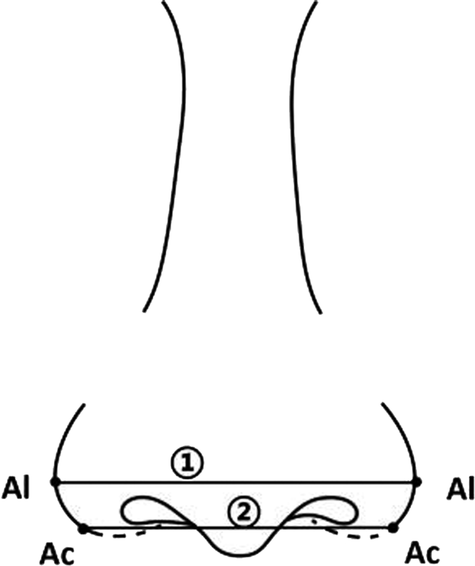
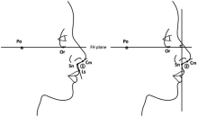
图2
鼻部侧面观示意图 Po:耳点;Or:眶下缘点;FH plane:眶耳平面;Sn:鼻下点;Cm:鼻小柱前点;Ls:上唇缘点;①鼻唇角(nasolabial angle,NLA);②鼻小柱倾角(columellar-facial angle,CFA)"

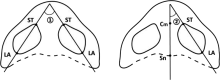
图3
鼻部仰面观示意图 ST:软三角点,即鼻孔内侧脚穹窿最高点;LA:鼻翼外侧脚近基底处最低点;Cm:鼻小柱前点;Sn:鼻下点ST-LA:鼻孔长轴径向;①两侧鼻孔长轴径向夹角;②鼻孔长轴径向与鼻小柱长轴的夹角"

| [1] |
Heiman AJ, Nair L, Kanth A, et al. Defining regional variation in nasal anatomy to guide ethnic rhinoplasty:A systematic review[J]. J Plast Reconstr Aesthetic Surg, 2022, 75(8):2784-2795.
doi: 10.1016/j.bjps.2022.04.058 |
| [2] |
Worasakwutiphong S, Chuang YF, Chang HW, et al. Nasal changes after orthognathic surgery for patients with prognathism and Class Ⅲ malocclusion:Analysis using three-dimensional photogrammetry[J]. J Formos Med Assoc, 2015, 114(2):112-123.
doi: 10.1016/j.jfma.2014.10.003 |
| [3] |
Hellak AF, Kirsten B, Schauseil M, et al. Influence of maxillary advancement surgery on skeletal and soft-tissue changes in the nose—a retrospective cone-beam computed tomography study[J]. Head Face Med, 2015, 11(1):23.
doi: 10.1186/s13005-015-0080-y |
| [4] |
Allar ML, Movahed R, Wolford LM, et al. Nasolabial changes following double jaw surgery[J]. J Craniofac Surg, 2019, 30(8):2560-2564.
doi: 10.1097/SCS.0000000000005876 pmid: 31689731 |
| [5] |
Rohrich RJ, Malafa MM, Ahmad J, et al. Managing alar flare in rhinoplasty[J]. Plast Reconstr Surg, 2017, 140(5):910-919.
doi: 10.1097/PRS.0000000000003786 pmid: 29068925 |
| [6] |
Moon KC, Han SK. Surgical anatomy of the Asian nose[J]. Facial Plast Surg Clin North Am, 2018, 26(3):259-268.
doi: 10.1016/j.fsc.2018.03.001 |
| [7] |
Choi SY, Kim SJ, Lee HY, et al. Esthetic nasolabial angle according to the degree of upper lip protrusion in an Asian population[J]. Am J Rhinol Allergy, 2018, 32(1):66-70.
doi: 10.2500/ajra.2018.32.4485 pmid: 29336294 |
| [8] |
Ahmed O, Dhinsa A, Popenko N, et al. Population-based assessment of currently proposed ideals of nasal tip projection and rotation in young women[J]. JAMA Facial Plast Surg, 2014, 16(5):310-318.
doi: 10.1001/jamafacial.2014.228 |
| [9] |
Ganske IM, Tan RA, Langa OC, et al. Does the nostril shape change after le fort I advancement in patients with unilateral complete cleft lip?[J]. J Oral Maxillofac Surg, 2020, 78(6):998-1005.
doi: 10.1016/j.joms.2020.01.010 |
| [10] | Suzen M, Dilaver E, Ak KB, et al. Analysis of inferior nasal morphology and nostrils following le fort I osteotomy[J]. J CraniofacSurg, 2022, 33(8):2682-2687. |
| [11] | Dindaroğlu F, Kutlu P, Duran GS, et al. Accuracy and reliability of 3D stereophotogrammetry:A comparison to direct anthropometry and 2D photogrammetry[J]. Angle Orthod, 2016, 86(3):487-494. |
| [12] | Atakan A, Özçırpıcı AA. Correlation between cephalometric nasal changes and patients’ perception after orthognathic surgery[J]. Am J Orthod Dentofac Orthop, 2021, 159(6):e449-e460. |
| [13] |
Baysal A, Sahan AO, Ozturk MA, et al. Reproducibility and reliability of three-dimensional soft tissue landmark identification using three-dimensional stereophotogrammetry[J]. Angle Orthod, 2016, 86(6):1004-1009.
pmid: 27023408 |
| [14] |
Guntaka PK, Kiang K, Caprio R, et al. Do patients treated with Invisalign have less swelling after orthognathic surgery than those with fixed orthodontic appliances?[J]. Am J Orthod Dentofac Orthop, 2023, 163(2):243-251.
doi: 10.1016/j.ajodo.2021.11.015 |
| [15] |
Coban G, Yavuz I, Karadas B, et al. Three-dimensional assess-ment of nasal changes after maxillary advancement with impaction using stereophotogrammetry[J]. Korean J Orthod, 2020, 50(4):249-257.
doi: 10.4041/kjod.2020.50.4.249 |
| [16] |
Almukhtar A, Khambay B, Ju X, et al. Comprehensive analysis of soft tissue changes in response to orthognathic surgery:Mandibular versus bimaxillary advancement[J]. Int J Oral Maxillofac Surg, 2018, 47(6):732-737.
doi: 10.1016/j.ijom.2017.11.014 |
| [17] |
Jung J, Lee CH, Lee JW, et al. Three dimensional evaluation of soft tissue after orthognathic surgery[J]. Head Face Med, 2018, 14(1):21.
doi: 10.1186/s13005-018-0179-z pmid: 30290762 |
| [18] |
D’Ettorre G, Farronato M, Candida E, et al. A comparison between stereophotogrammetry and smartphone structured light technology for three-dimensional face scanning[J]. Angle Orthod, 2022, 92(3):358-363.
doi: 10.2319/040921-290.1 pmid: 35015071 |
| [19] |
Pan FW, Liu JL, Cen YY, et al. Accuracy of RGB-D camera-based and stereophotogrammetric facial scanners:A comparative study[J]. J Dent, 2022, 127:104302.
doi: 10.1016/j.jdent.2022.104302 |
| [20] | Zhao YJ, Xiong YX, Wang Y. Three-dimensional accuracy of facial scan for facial deformities in clinics:A new evaluation method for facial scanner accuracy[J]. PLoS One, 2017, 12(1):e0169402. |
| [21] |
ten Harkel TC, Vinayahalingam S, Ingels KJAO, et al. Reliability and agreement of 3D anthropometric measurements in facial palsy patients using a low-cost 4D imaging system[J]. IEEE Trans Neural Syst Rehabil Eng, 2020, 28(8):1817-1824.
doi: 10.1109/TNSRE.7333 |
| [22] |
Petrides G, Clark JR, Low H, et al. Three-dimensional scanners for soft-tissue facial assessment in clinical practice[J]. J Plast Reconstr Aesthetic Surg, 2021, 74(3):605-614.
doi: 10.1016/j.bjps.2020.08.050 |
| [23] |
Chen ZC, Albdour MN, Lizardo JA, et al. Precision of three-dimensional stereo-photogrammetry (3dMDTM) in anthropometry of the auricle and its application in microtiare construction[J]. J Plast Reconstr Aesthetic Surg, 2015, 68(5):622-631.
doi: 10.1016/j.bjps.2015.02.020 |
| [24] |
Raffone C, Gianfreda F, Pompeo MG, et al. Chairside virtual patient protocol. Part 2:Management of multiple face scans and alignment predictability[J]. J Dent, 2022, 122:104123.
doi: 10.1016/j.jdent.2022.104123 |
| [25] |
Thurzo A, Strunga M, Havlínová R, et al. Smartphone-based facial scanning as a viable tool for facially driven orthodontics?[J]. Sensors (Basel), 2022, 22(20):7752.
doi: 10.3390/s22207752 |
| [26] |
Liu JL, Zhang CH, Cai RL, et al. Accuracy of 3-dimensional stereophotogrammetry: Comparison of the 3dMD and Bellus3D facial scanning systems with one another and with direct anthropometry[J]. Am J Orthod Dentofacial Orthop, 2021, 160(6):862-871.
doi: 10.1016/j.ajodo.2021.04.020 |
| [27] |
Seon S, Lee HW, Jeong BJ, et al. Study of soft tissue changes in the upper lip and nose after backward movement of the maxilla in orthognathic surgery[J]. J Korean Assoc Oral Maxillofac Surg, 2020, 46(6):385-392.
doi: 10.5125/jkaoms.2020.46.6.385 pmid: 33377463 |
| [28] |
Lee JY, Kim YI, Hwang DS, et al. Effect of setback Le Fort I osteotomy on midfacial soft-tissue changes as evaluated by cone-beam computed tomography superim position for cases of skeletal Class Ⅲ malocclusion[J]. Int J Oral Maxillofac Surg, 2013, 42(6):790-795.
doi: 10.1016/j.ijom.2012.11.012 |
| [29] | Sabri H, Tehranchi A, Sarkarat F. 3-dimensional analysis of nasal soft tissue alterations following maxillary Lefort I advancement with and without impaction using 3D photogrammetry scanner[J]. Oral Maxillofac Surg, 2022:1-13. |
| [30] |
Denadai R, Chou PY, Yao CF, et al. Effect of le fort I maxillary repositioning on three-dimensional nasal tip rotation:A comparative study with implication for the Asian nose[J]. Plast Reconstr Surg, 2021, 147(4):903-914.
doi: 10.1097/PRS.0000000000007774 pmid: 33750094 |
| [31] |
Denadai R, Chou PY, Lin YY, et al. Type of maxillary segment mobilization affects three-dimensional nasal morphology[J]. J Plast Reconstr Aesthetic Surg, 2021, 74(3):592-604.
doi: 10.1016/j.bjps.2020.08.119 |
| [32] |
de Sousa Gil AP, Guijarro-Martínez R, Haas OL, et al. Three-dimensional analysis of nasolabial soft tissue changes after Le Fort I osteotomy:A systematic review of the literature[J]. Int J Oral Maxillofac Surg, 2019, 48(9):1185-1200.
doi: 10.1016/j.ijom.2019.01.028 |
| [33] |
Üstün GG, Konaş E, El H, et al. The effects of maxillary movements on nasal aesthetics following orthognathic surgery[J]. J Craniofac Surg, 2020, 31(3):796-800.
doi: 10.1097/SCS.0000000000006167 pmid: 31934978 |
| [34] |
Sawh-Martinez R, Lin AM, DeSesa CR, et al. Clockwise and counterclockwise le fort I movements influence nasolabial morphology differently[J]. Plast Reconstr Surg, 2018, 142(6):1572-1581.
doi: 10.1097/PRS.0000000000004988 pmid: 30188468 |
| [35] |
Schendel SA, Williamson LW. Muscle reorientation following superior repositioning of the maxilla[J]. J Oral Maxillofac Surg, 1983, 41(4):235.
doi: 10.1016/0278-2391(83)90265-3 |
| [36] |
Khamashta-Ledezma L, Naini FB. Prospective assessment of maxillary advancement effects:Maxillary incisor exposure, and upper lip and nasal changes[J]. Am J Orthod Dentofac Orthop, 2015, 147(4):454-464.
doi: 10.1016/j.ajodo.2014.11.028 |
| [37] |
Westermark AH, Bystedt H, von Konow L, et al. Nasolabial morphology after le fort I osteotomies effect of alar base suture[J]. Int J Oral Maxillofac Surg, 1991, 20(1):25-30.
doi: 10.1016/S0901-5027(05)80690-3 |
| [38] |
Muradin MSM, Rosenberg A, van der Bilt A, et al. The effect of alar cinch sutures and V-Y closure on soft tissue dynamics after Le Fort I intrusion osteotomies[J]. J Cranio Maxillofac Surg, 2009, 37(6):334-340.
doi: 10.1016/j.jcms.2009.03.004 |
| [39] | Monnazzi MS, Mannarino FS, Gabrielli MFR. Extraoral alar base cinch. A modification for the technique[J]. J Oral Maxillofac Surg Med Pathol, 2014, 26(2):142-144. |
| [40] |
Mani V, Panicker P, Shenoy A, et al. Evaluation of changes in the alar base width following lefort 1 and AMO with conventional alar cinch suturing:A photographic study of 100 cases[J]. J Maxillofac Oral Surg, 2020, 19(1):21-25.
doi: 10.1007/s12663-019-01227-8 |
| [41] |
Raithatha R, Naini FB, Patel S, et al. Long-term stability of limiting nasal alar base width changes with a cinch suture following Le Fort I osteotomy with submentalintubation[J]. Int J Oral Maxillofac Surg, 2017, 46(11):1372-1379.
doi: 10.1016/j.ijom.2017.04.027 |
| [42] |
Mahsoub R, Naini FB, Patel S, et al. Nasolabial angle and nasal tip elevation changes in profile view following a Le Fort I osteotomy with or without the use of an alar base cinch suture:A long-term cohort study[J]. Oral Surg Oral Med Oral Pathol Oral Radiol, 2020, 130(4):379-386.
doi: 10.1016/j.oooo.2020.05.011 |
| [43] |
Ishida T, Manabe A, Yang SS, et al. An orthodontic-orthognathic patient with obstructive sleep apnea treated with Le Fort I osteotomy advancement and alar cinch suture combined with a muco-musculo-periosteal V-Y closure to minimize nose deformity[J]. Angle Orthod, 2019, 89(6):946-952.
doi: 10.2319/052818-406.1 pmid: 30698453 |
| [44] | Yang J, Chu Y, Liao H, et al. Comparison of the effectiveness between conventional and modified cinch suture techniques in LeFort I osteotomy:A systematic review and meta-analysis of randomized control trials[J]. Ann Plast Surge, 2023,doi:10.1097/SAP.0000000000003354. |
| [45] |
Gil APS, Machado-Fernández A, Guijarro-Martínez R, et al. Le Fort I osteotomy and soft tissue response:A retrospective cohort study comparing three different techniques[J]. J Cranio Maxillofac Surg, 2022, 50(2):107-113.
doi: 10.1016/j.jcms.2021.11.009 |
| [46] |
Cho YS, Hwang KG, Park CJ. Postoperative effects of anterior nasal spine bone harvesting on overall nasal shape[J]. Clin Oral Implants Res, 2013, 24(6):618-622.
doi: 10.1111/clr.2013.24.issue-6 |
| [47] |
Jung S, Kim JY, Jung YS, et al. Reliability of anterior nasal spine as a reference point after LeFort I surgery using three-dimensional analysis[J]. J Craniofac Surg, 2022, 33(7):2104-2108.
doi: 10.1097/SCS.0000000000008619 pmid: 35261362 |
| [48] |
Zhong YH, Zhu YJ, Jiang TR, et al. A novel study on alar mobility of HAN female by 3dMD dynamic surface imaging system[J]. Aesth Plast Surg, 2022, 46(1):364-372.
doi: 10.1007/s00266-021-02386-1 |
| [1] | 石慧, 陆胜男, 夏文倩, 章婷, 贡敏, 梅予锋. 螺旋器分裂基托式矫治装置远移上颌第一磨牙的三维有限元分析[J]. 口腔医学, 2023, 43(11): 1002-1007. |
| [2] | 周志捷, 洪越扬, 许衍. 下颌第二磨牙近中阻生的局部牙列及颌骨形态的三维分析[J]. 口腔医学, 2023, 43(11): 1008-1013. |
| [3] | 魏晓渝, 张冠凝, 赵青. 正畸诱导炎性牙根吸收影响因素的研究进展[J]. 口腔医学, 2023, 43(11): 1034-1040. |
| [4] | 胡洁琼, 李青奕. 三维立体摄影测量技术与正畸[J]. 口腔医学, 2023, 43(11): 1047-1052. |
| [5] | 林祥祥, 余飞, 宋艺蔚, 王莞, 弓国梁, 林新平. 内收上颌切牙时唇腭侧牙槽骨改建影响因素的研究进展[J]. 口腔医学, 2023, 43(11): 1053-1056. |
| [6] | 周佳琦, 吴栋, 吴樱仪, 周力, 李宇. 双牙合板式透明矫治器作用力值的测量及其影响因素分析[J]. 口腔医学, 2023, 43(10): 894-898. |
| [7] | 许嘉宁, 金作林, 刘佳. 3D打印在口腔正畸领域的应用进展[J]. 口腔医学, 2023, 43(10): 925-929. |
| [8] | 周妍, 彭友俭. 双矩形附件对透明矫治器磨牙近中移动的三维有限元分析[J]. 口腔医学, 2023, 43(10): 883-888. |
| [9] | 康芙嘉, 张茜雅, 余磊, 王宋庆, 张皓岩, 李欣怡, 朱宪春. 微种植体辅助无托槽隐形矫治器压低上颌前牙的三维有限元分析[J]. 口腔医学, 2023, 43(9): 796-802. |
| [10] | 马晓晴, 孙建锋, 叶茂, 邢允波, 邓学韬, 朱梦娇, 许衍. 不等抗力偶直立下颌高位近中阻生第二磨牙的临床研究[J]. 口腔医学, 2023, 43(9): 803-807. |
| [11] | 蒋镇泽, 邹明媛, 弓国梁, 林新平. 第二磨牙在不同情况下对改良C型腭板远中移动上颌磨牙的影响——三维有限元分析[J]. 口腔医学, 2023, 43(9): 808-813. |
| [12] | 马燕, 朱道俣, 张春霞, 陶李明. 成人骨性Ⅱ类错𬌗矫治前后上颌前牙牙根吸收的研究[J]. 口腔医学, 2023, 43(9): 814-818. |
| [13] | 陈燕玲, 张晟, 麦理想, 欧卜宾. 成人安氏Ⅱ类拔牙病例无托槽隐形矫治的支抗控制效果评估[J]. 口腔医学, 2023, 43(9): 819-824. |
| [14] | 黄钖钖, 刘浩, 王诗语, 范典, 王鹏来, 袁长永. 过矫治设计在无托槽隐形矫治中的应用现状[J]. 口腔医学, 2023, 43(9): 849-853. |
| [15] | 陈云, 曹灵, 王雅欣, 于剑南. 815颗儿童上颌多生牙的回顾性分析[J]. 口腔医学, 2023, 43(8): 717-721. |
| 阅读次数 | ||||||
|
全文 |
|
|||||
|
摘要 |
|
|||||
 苏公网安备32010602011670号