| [1] |
Wiegand S, Dietz A. Vascular malformations of the head and neck[J]. Laryngorhinootologie, 2021, 100(1): 65-76.
|
| [2] |
Markovic JN, Shortell CK. Venous malformations[J]. J Cardiovasc Surg, 2021, 62(5): 456-466.
|
| [3] |
Cooke-Barber J, Kreimer S, Patel M, et al. Venous malformations[J]. Semin Pediatr Surg, 2020, 29(5): 150976.
doi: 10.1016/j.sempedsurg.2020.150976
pmid: 33069284
|
| [4] |
Chen S, Wang Y, Kong LL, et al. Role of UDP-glucose ceramide glucosyltransferase in venous malformation[J]. Front Cell Dev Biol, 2023, 11: 1178045.
|
| [5] |
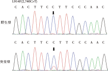
施磊, 孙圆圆, 甘露, 等. 西罗莫司对TIE2-L914F突变导致的静脉畸形血管内皮细胞增殖和凋亡的影响及其体外机制探究[J]. 安徽医科大学学报, 2023, 58(4): 561-567.
|
| [6] |
Anisimov A, Fang ST, Hemanthakumar KA, et al. The angiopoietin receptor Tie2 is atheroprotective in arterial endothelium[J]. Nat Cardiovasc Res, 2023, 2(3): 307-321.
|
| [7] |
Eklund L, Olsen BR. Tie receptors and their angiopoietin ligands are context-dependent regulators of vascular remodeling[J]. Exp Cell Res, 2006, 312(5): 630-641.
doi: 10.1016/j.yexcr.2005.09.002
pmid: 16225862
|
| [8] |
Goines J, Boscolo E. A xenograft model for venous malformation[J]. Methods Mol Biol, 2021, 2206: 179-192.
doi: 10.1007/978-1-0716-0916-3_13
pmid: 32754818
|
| [9] |
Limaye N, Boon LM, Vikkula M. From germline towards somatic mutations in the pathophysiology of vascular anomalies[J]. Hum Mol Genet, 2009, 18(R1): R65-R74.
|
| [10] |
Teichert M, Milde L, Holm A, et al. Pericyte-expressed Tie2 controls angiogenesis and vessel maturation[J]. Nat Commun, 2017, 8: 16106.
doi: 10.1038/ncomms16106
pmid: 28719590
|
| [11] |
Zhang YC, Liu JQ, Zou T, et al. DPSCs treated by TGF-β1 regulate angiogenic sprouting of three-dimensionally co-cultured HUVECs and DPSCs through VEGF-Ang-Tie2 signaling[J]. Stem Cell Res Ther, 2021, 12(1): 281.
doi: 10.1186/s13287-021-02349-y
pmid: 33971955
|
| [12] |
Nätynki M, Kangas J, Miinalainen I, et al. Common and specific effects of TIE2 mutations causing venous malformations[J]. Hum Mol Genet, 2015, 24(22): 6374-6389.
doi: 10.1093/hmg/ddv349
pmid: 26319232
|
| [13] |
Xu M, Chen XL, Chen DW, et al. FoxO1: A novel insight into its molecular mechanisms in the regulation of skeletal muscle differentiation and fiber type specification[J]. Oncotarget, 2017, 8(6): 10662-10674.
doi: 10.18632/oncotarget.12891
pmid: 27793012
|
| [14] |
Zhao T, Wang J, He AX, et al. Mebhydrolin ameliorates glucose homeostasis in type 2 diabetic mice by functioning as a selective FXR antagonist[J]. Metabolism, 2021, 119: 154771.
|
| [15] |
Deng H, Gong YX, Chen Y, et al. Porphyromonas gingivalis lipopolysaccharide affects the angiogenic function of endothelial progenitor cells via Akt/FoxO1 signaling[J]. J Periodontal Res, 2022, 57(4): 859-868.
doi: 10.1111/jre.13024
pmid: 35694806
|
| [16] |
Ke J, Wei R, Yu F, et al. Liraglutide restores angiogenesis in palmitate-impaired human endothelial cells through PI3K/Akt-Foxo1-GTPCH1 pathway[J]. Peptides, 2016, 86: 95-101.
doi: S0196-9781(16)30212-1
pmid: 27777063
|
| [17] |
王肖, 招洛丹, 彭志翔. 胰岛素样生长因子1经 PI3K/Akt 信号通道对人牙髓细胞凋亡的抑制调控[J]. 口腔医学, 2014, 34(9): 674-678.
|
| [18] |
Liu ZK, Liu HH, Liu SB, et al. SIRT1 activation promotes bone repair by enhancing the coupling of type H vessel formation and osteogenesis[J]. Cell Prolif, 2024, 57(6): e13596.
|
| [19] |
di Pietro M, Scotti L, Irusta G, et al. Local administration of platelet-derived growth factor B(PDGFB)improves follicular development and ovarian angiogenesis in a rat model of Polycystic Ovary Syndrome[J]. Mol Cell Endocrinol, 2016, 433: 47-55.
|
| [20] |
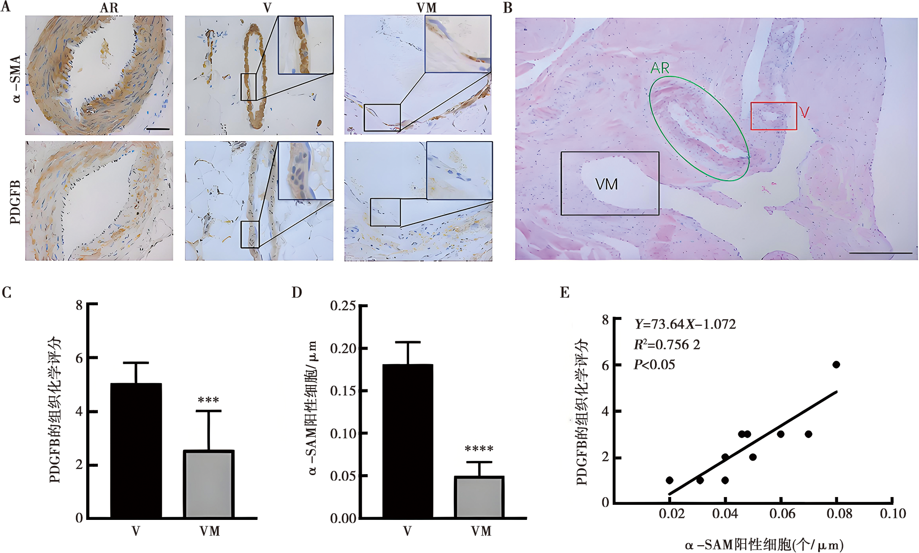
Lin YZ, Gahn J, Banerjee K, et al. Role of endothelial PDGFB in arterio-venous malformations pathogenesis[J]. Angiogenesis, 2024, 27(2): 193-209.
|
| [21] |
Mammoto A, Hendee K, Muyleart M, et al. Endothelial Twist1-PDGFB signaling mediates hypoxia-induced proliferation and migration of αSMA-positive cells[J]. Sci Rep, 2020, 10(1): 7563.
doi: 10.1038/s41598-020-64298-5
pmid: 32371931
|

 )
)
 苏公网安备32010602011670号