
口腔医学 ›› 2023, Vol. 43 ›› Issue (12): 1145-1152.doi: 10.13591/j.cnki.kqyx.2023.12.017
• 综述 • 上一篇
汤佩,吴建勇( )
)
修回日期:2023-01-08
出版日期:2023-12-28
发布日期:2023-12-28
通讯作者:
吴建勇
E-mail:wujianyong@xinhuamed.com.cn
基金资助:
TANG Pei,WU Jianyong()
Revised:2023-01-08
Online:2023-12-28
Published:2023-12-28
Contact:
WU Jianyong
E-mail:wujianyong@xinhuamed.com.cn
摘要:
牙弓和基骨的大小和形态现已成为正畸医生对错牙合畸形诊断、方案设计及疗效评估的重要参数,明确不同错牙合畸形患者牙弓和基骨的横向特征以及影响因素,有利于正畸医生对治疗的把控。因此,本文就牙弓和基骨相关测量方法,不同错牙合畸形患者牙弓及基骨横向特征及影响因素作一综述。
中图分类号:
汤佩,吴建勇. 牙弓及基骨横向特征的研究进展[J]. 口腔医学, 2023, 43(12): 1145-1152.
TANG Pei,WU Jianyong. Research progress of transverse characteristics of the dental arch and basal bone[J]. Stomatology, 2023, 43(12): 1145-1152.

图1
使用PAC测量上、下颌骨宽度及上、下牙弓宽度 U6:上颌第一磨牙最颊侧点;L6:下颌第一磨牙最颊侧点;J:上颌结节与颧突支柱交点;Ag:下颌双侧角前切迹点"
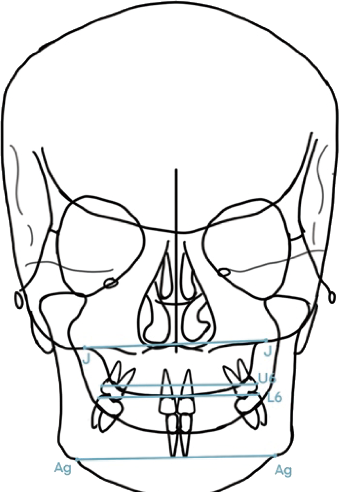
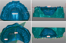
图2
模型测量法 WALA:WALA嵴点; B:牙冠最颊侧点; C:牙尖点; F:中央窝点; L:牙冠最舌侧点; Fa:临床牙冠面轴线的中点;平面1:牙合平面;平面2:上颌第一磨牙近远中腭尖与舌沟所形成平面;平面3:正中矢状面;∠1:过Fa点的切线与牙合平面所成的角度;∠2:平面2与平面3之间的夹角;∠3:Wilson曲线与参考牙合平面所形成的角度"

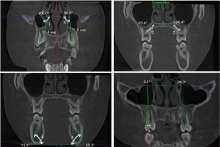
图3
CBCT测量法 ∠1:牙长轴与FH平面(右侧耳点与双侧眶点所形成的平面)所形成的角度即∠3;∠2:牙长轴与腭平面所形成的角度;∠3:牙长轴与双侧眶下缘最低点连线所形成的角度;∠4:牙长轴与下颌平面所形成的角度;∠5:牙长轴与正中矢状面所形成的角度"
| [1] |
Goldstein MS. Changes in dimensions and form of the face and head with age[J]. Am J Phys Anthropol, 1936, 22(1):37-89.
doi: 10.1002/ajpa.v22:1.o |
| [2] |
Hwang S, Jeong S, Choi YJ, et al. Three-dimensional evaluation of dentofacial transverse widths of adults with various vertical facial patterns[J]. Am J Orthod Dentofacial Orthop, 2018, 153(5):692-700.
doi: 10.1016/j.ajodo.2017.08.026 |
| [3] | Zhang X, He JM, Zheng WY. Comparison of rapid maxillary expansion and pre-fabricated myofunctional appliance for the management of mouth breathers with Class Ⅱ malocclusion[J]. Eur Rev Med Pharmacol Sci, 2021, 25(1):16-23. |
| [4] |
Lundström AF. Malocclusion of the teeth regarded as a problem in connection with the apical base[J]. Int J Orthod Oral Surg Radiogr, 1925, 11(11):1022-1042.
doi: 10.1016/S0099-6963(25)80005-X |
| [5] | 皮昕. 口腔解剖生理学[M]. 6版. 北京: 人民卫生出版社, 2007. |
| [6] |
Howes AE. Model analysis for treatment planning[J]. Am J Orthod, 1952, 38(3):183-207.
doi: 10.1016/0002-9416(52)90107-3 |
| [7] | Andrews L. The six elements of orofacial harmony[J]. Andrews J, 2000, 1(1):13-22. |
| [8] |
Kong-Zárate CY, Carruitero MJ, Andrews WA. Distances between mandibular posterior teeth and the WALA ridge in Peruvians with normal occlusion[J]. Dental Press J Orthod, 2017, 22(6):56-60.
doi: S2176-94512017000600056 pmid: 29364380 |
| [9] |
Sampermans G, Sawaljanow A, Proff P, et al. Ideal transverse position of mandibular first molars based on CBCT-derived alveolar bone coverage[J]. Ann Anat, 2022, 241: 151908.
doi: 10.1016/j.aanat.2022.151908 |
| [10] |
Yoon S, Lee DY, Jung SK. Influence of changing various parameters in miniscrew-assisted rapid palatal expansion: A three-dimensional finite element analysis[J]. Korean J Orthod, 2019, 49(3):150-160.
doi: 10.4041/kjod.2019.49.3.150 pmid: 31149605 |
| [11] |
Andrews LF. The 6-elements orthodontic philosophy: Treatment goals, classification, and rules for treating[J]. Am J Orthod Dentofacial Orthop, 2015, 148(6):883-887.
doi: 10.1016/j.ajodo.2015.09.011 |
| [12] |
Lorente C, Lorente P, Perez-Vela M, et al. Quad-helix compression to decompensate molar inclination prior to skeletal expansion[J]. J Orofac Orthop, 2020, 81(2):142-149.
doi: 10.1007/s00056-019-00212-7 pmid: 32006047 |
| [13] |
Araujo E, Tanaka OM. The brodie bite: Addressing a confounding orthodontic problem[J]. AJO DO Clin Companion, 2021, 1(4):232-244.
doi: 10.1016/j.xaor.2021.10.002 |
| [14] | Pont A. Der zahn-index in der orthodontie[J]. Zahnartzliche Orthopadie, 1909, 3: 306-321. |
| [15] |
Dalidjan M, Sampson W, Townsend G. Prediction of dental arch development: An assessment of Pont’s Index in three human populations[J]. Am J Orthod Dentofacial Orthop, 1995, 107(5):465-475.
doi: 10.1016/S0889-5406(95)70113-3 |
| [16] |
Ricketts RM. Perspectives in the clinical application of cephalometrics. The first fifty years[J]. Angle Orthod, 1981, 51(2):115-150.
doi: 10.1043/0003-3219(1981)051<0115:PITCAO>2.0.CO;2 pmid: 6942666 |
| [17] |
Kohakura S, Kasai K, Ohno I, et al. Relationship between maxillofacial morphology and morphological characteristics of vertical sections of the mandible obtained by CT scanning[J]. J Nihon Univ Sch Dent, 1997, 39(2):71-77.
pmid: 9293703 |
| [18] |
Schulze RW, Drage NA. Cone-beam computed tomography and its applications in dental and maxillofacial radiology[J]. Clin Radiol, 2020, 75(9):647-657.
doi: S0009-9260(20)30158-6 pmid: 32451060 |
| [19] |
Dindaroğlu F, Duran GS, Tekeli A, et al. Evaluation of the relationship between curve of spee, WALA-FA distance and curve of Wilson in normal occlusion[J]. Turk J Orthod, 2016, 29(4):91-97.
doi: 10.5152/TurkJOrthod. |
| [20] |
Škrinjarić A, Šlaj M, Šlaj M. Fluctuating dental arch asymmetry in different malocclusion groups[J]. Acta Stomatol Croat, 2018, 52(2):105-113.
doi: 10.15644/asc52/2/3 pmid: 30034009 |
| [21] |
Kato M, Arai K. Relationship between dental and basal arch forms in mandibular anterior crowding[J]. Am J Orthod Dentofacial Orthop, 2022, 161(1):53-64.
doi: 10.1016/j.ajodo.2020.06.046 |
| [22] |
Fu KY, Fang S, Fan XF, et al. Analysis of dental and basal bone arch form correlations in skeletal Class Ⅱmalocclusion[J]. Am J Orthod Dentofacial Orthop, 2021, 159(2):202-209.e2.
doi: 10.1016/j.ajodo.2019.12.026 |
| [23] |
Ferreira MC, Freitas KMS, Herrera-Sanches FS, et al. Evaluation of mandibular first molars’ axial inclination and alveolar morphology in different facial patterns: A CBCT study[J]. Eur J Dent, 2020, 14(2):250-259.
doi: 10.1055/s-0040-1709932 pmid: 32438429 |
| [24] |
Moon HW, Nam W, AhnHW, et al. Development of a maxillomandibular arch form based on the center of resistance of teeth using cone-beam computed tomography[J]. Am J Orthod Dentofacial Orthop, 2022, 161(2):208-219.
doi: 10.1016/j.ajodo.2020.07.041 |
| [25] |
Al-Mashraqi AA, Alhammadi MS, Gadi AA, et al. Accuracy and reproducibility of permanent dentitions and dental arch measurements: Comparing three different digital models with a plaster study cast[J]. Int J Comput Dent, 2021, 24(4):353-362.
pmid: 34931771 |
| [26] | 郝俊玲. 基于基骨的个性化下颌牙弓形态研究[D]. 福州: 福建医科大学, 2018. |
| [27] |
Shokri A, Miresmaeili A, Farhadian N, et al. Effect of changing the head position on accuracy of transverse measurements of the maxillofacial region made on cone beam computed tomography and conventional posterior-anterior cephalograms[J]. Dentomaxillofac Radiol, 2017, 46(5):20160180.
doi: 10.1259/dmfr.20160180 |
| [28] |
De Grauwe A, Ayaz I, Shujaat S, et al. CBCT in orthodontics: A systematic review on justification of CBCT in a paediatric population prior to orthodontic treatment[J]. Eur J Orthod, 2019, 41(4):381-389.
doi: 10.1093/ejo/cjy066 |
| [29] |
Prasad M, Kannampallil ST, Talapaneni AK, et al. Evaluation of arch width variations among different skeletal patterns in South Indian population[J]. J Nat Sci Biol Med, 2013, 4(1):94-102.
doi: 10.4103/0976-9668.107267 |
| [30] |
Shafique HZ, Zaheer R, Jan A, et al. Comparison of tooth widths, arch widths and arch lengths in Class-Ⅰ normal dentition to Class-Ⅰ and Ⅱ crowded dentitions[J]. Pak J Med Sci, 2021, 37(2):345-350.
doi: 10.12669/pjms.37.2.3240 pmid: 33679911 |
| [31] |
Lee SJ, Lee S, Lim J, et al. Method to classify dental arch forms[J]. Am J Orthod Dentofacial Orthop, 2011, 140(1):87-96.
doi: 10.1016/j.ajodo.2011.03.016 |
| [32] | Yang DP, Liang SR, Zhang K, et al. Evaluation of growth and development of late mixed dentitionupper dental arch with normal occlusion using 3-dimensional digital models[J]. J Healthc Eng, 2019, 2019: 4191848. |
| [33] |
Al-Hilal LH, Sultan K, Hajeer MY, et al. An evaluation of mandibular dental and basal arch dimensions in Class Ⅰ and Class Ⅱ division 1 adult Syrian patients using cone-beam computed tomography[J]. J Contemp Dent Pract, 2018, 19(4):431-437.
pmid: 29728549 |
| [34] |
Baka ZM, Akin M, Ucar FI, et al. Cone-beam computed tomography evaluation of dentoskeletal changes after asymmetric rapid maxillary expansion[J]. Am J Orthod Dentofacial Orthop, 2015, 147(1):61-71.
doi: 10.1016/j.ajodo.2014.09.014 |
| [35] |
Son WS, Oh S, Kim YI, et al. Comparison of maxillary basal arch forms using the root apex in adult women with different skeletal patterns: A pilot study[J]. Am J Orthod Dentofacial Orthop, 2020, 158(4):572-578.
doi: 10.1016/j.ajodo.2019.09.021 |
| [36] |
Crossley AM, Campbell PM, Tadlock LP, et al. Is there a relationship between dental crowding and the size of the maxillary or mandibular apical base?[J]. Angle Orthod, 2020, 90(2):216-223.
doi: 10.2319/051019-324.1 pmid: 31549856 |
| [37] | Golshah A, Rezaei N, Heshmati S. Buccolingualinclination of canine and first and second molar teeth and the curve of Wilson in different sagittal skeletal patterns of adults using cone-beam computed tomography[J]. Int J Dent, 2020, 2020: 8893778. |
| [38] |
Hwang S, Song J, Lee J, et al. Three-dimensional evaluation of dentofacial transverse widths in adults with different sagittal facial patterns[J]. Am J Orthod Dentofacial Orthop, 2018, 154(3):365-374.
doi: 10.1016/j.ajodo.2017.11.041 |
| [39] |
Al-Zubair NM. Dental arch asymmetry[J]. Eur J Dent, 2014, 8(2):224-228.
doi: 10.4103/1305-7456.130608 pmid: 24966774 |
| [40] | Patel D, Mehta F, Patel N, et al. Evaluation of arch width among Class Ⅰ normal occlusion, Class Ⅱ Division 1, Class Ⅱ Division 2, and Class Ⅲ malocclusion in Indian population[J]. Contemp Clin Dent, 2015, 6(Suppl 1):S202-S209. |
| [41] |
Buschang PH, Stroud J, Alexander RG. Differences in dental arch morphology among adult females with untreated Class Ⅰ and Class Ⅱ malocclusion[J]. Eur J Orthod, 1994, 16(1):47-52.
doi: 10.1093/ejo/16.1.47 |
| [42] |
曹正飞, 王青青, 关慧娟, 等. 安氏Ⅱ类1分类与安氏Ⅰ类错牙合畸形患者牙弓宽度比较的Meta分析[J]. 中国实用口腔科杂志, 2019, 12(1):36-41.
doi: 10.19538/j.kq.2019.01.008 |
| [43] |
Shu R, Han XL, Wang YT, et al. Comparison of arch width, alveolar width and buccolingual inclination of teeth between Class Ⅱ division 1 malocclusion and Class Ⅰ occlusion[J]. Angle Orthod, 2013, 83(2):246-252.
doi: 10.2319/052412-427.2 |
| [44] |
Shewinvanakitkul W, Hans MG, Narendran S, et al. Measuring buccolingual inclination of mandibular canines and first molars using CBCT[J]. Orthod Craniofac Res, 2011, 14(3):168-174.
doi: 10.1111/j.1601-6343.2011.01518.x pmid: 21771272 |
| [45] | Shahroudi AS, Etezadi T. Correlation between dental arch width and sagittal dento-skeletal morphology in untreated adults[J]. J Dent(Tehran), 2013, 10(6):522-531. |
| [46] |
Ball RL, Miner RM, Will LA, et al. Comparison of dental and apical base arch forms in Class Ⅱ Division 1 and Class Ⅰ malocclusions[J]. Am J Orthod Dentofacial Orthop, 2010, 138(1):41-50.
doi: 10.1016/j.ajodo.2008.11.026 |
| [47] |
Dindaroğlu F, Duran GS, Aras I. Three-dimensional evaluation of morphologic tooth symmetry in various malocclusions[J]. Am J Orthod Dentofacial Orthop, 2016, 150(3):459-466.
doi: 10.1016/j.ajodo.2016.04.017 |
| [48] |
Slaj M, Spalj S, Pavlin D, et al. Dental archforms in dentoalveolarclass Ⅰ, Ⅱ and Ⅲ[J]. Angle Orthod, 2010, 80(5):919-924.
doi: 10.2319/112609-672.1 |
| [49] |
Suk KE, Park JH, Bayome M, et al. Comparison between dental and basal arch forms in normal occlusion and Class Ⅲ malocclusions utilizing cone-beam computed tomography[J]. Korean J Orthod, 2013, 43(1):15-22.
doi: 10.4041/kjod.2013.43.1.15 |
| [50] |
Chen YJ, Yao CC, Chang ZC, et al. Characterization of facial asymmetry in skeletal Class Ⅲ malocclusion and its implications for treatment[J]. Int J Oral Maxillofac Surg, 2019, 48(12):1533-1541.
doi: 10.1016/j.ijom.2019.06.014 |
| [51] |
Koo YJ, Choi SH, Keum BT, et al. Maxillomandibular arch width differences at estimated centers of resistance: Comparison between normal occlusion and skeletal Class Ⅲ malocclusion[J]. Korean J Orthod, 2017, 47(3):167.
doi: 10.4041/kjod.2017.47.3.167 |
| [52] |
Zou W, Wu JQ, Jiang JH, et al. Archform comparisons between skeletal Class Ⅱ and Ⅲ malocclusions[J]. PLoS One, 2014, 9(6):e100655.
doi: 10.1371/journal.pone.0100655 |
| [53] |
Ahn J, Kim SJ, Lee JY, et al. Transverse dental compensation in relation to sagittal and transverse skeletal discrepancies in skeletal Class Ⅲ patients[J]. Am J Orthod Dentofacial Orthop, 2017, 151(1):148-156.
doi: 10.1016/j.ajodo.2016.06.031 |
| [54] |
Forster CM, Sunga E, Chung CH. Relationship between dental arch width and vertical facial morphology in untreated adults[J]. Eur J Orthod, 2008, 30(3):288-294.
doi: 10.1093/ejo/cjm113 |
| [55] |
Grippaudo C, Oliva B, Greco AL, et al. Relationship between vertical facial patterns and dental arch form in Class Ⅱ malocclusion[J]. Prog Orthod, 2013, 14: 43.
doi: 10.1186/2196-1042-14-43 pmid: 24326093 |
| [56] | 刘晔, 沈悦, 张士珑, 等. 不同垂直骨面型安氏Ⅱ-1类错牙合畸形牙弓宽度分析[J]. 现代口腔医学杂志, 2020, 34(1):25-27, 24. |
| [57] | 金超超, 邵玶. Ⅲ类错牙合垂直骨面型与牙弓宽度关系的研究[J]. 口腔医学, 2018, 38(1):52-54. |
| [58] | Mohan S, Dusanapudi LN. Relationship of curve of Spee&dentofacial morphology in North Indian population-An in vitro study[J]. J Pierre Fauchard AcadIndia Sect, 2014, 28(2):61-67. |
| [59] | Hampe T, Krohn S, Schmitt F, et al. The variability of the curve of Spee: An analysis of multiple setups of the same Angle Class Ⅰ patient case[J]. J OrofacOrthop, 2020, 81(2):89-99. |
| [60] |
Paulino V, Paredes V, Gandia JL, et al. Prediction of arch length based on intercanine width[J]. Eur J Orthod, 2008, 30(3):295-298.
doi: 10.1093/ejo/cjm115 |
| [61] | Kareem FA, Rauf AM, Noori AJ, et al. Prediction of the dental arch perimeter in a Kurdish sample in sulaimanicity based on other linear dental arch measurements as a malocclusion preventive measure[J]. Comput Math Methods Med, 2020, 2020: 8869996. |
| [62] |
Al-Zubair NM. The relationship between mandibular arch length and widths in a sample of Yemeni subjects with normal dento-Skeletal relationship[J]. J Orthod Sci, 2013, 2(4):120-123.
doi: 10.4103/2278-0203.123198 pmid: 24987653 |
| [63] |
Bishara SE, Jakobsen JR, Treder J, et al. Arch width changes from 6 weeks to 45 years of age[J]. Am J Orthod Dentofacial Orthop, 1997, 111(4):401-409.
doi: 10.1016/S0889-5406(97)80022-4 |
| [64] |
Sayania B, Merchant M, Josephs P, et al. Changes in the buccolingual inclination of first molars with growth in untreated subjects: A longitudinal study[J]. Angle Orthod, 2017, 87(5):681-687.
doi: 10.2319/120716-878.1 pmid: 28481630 |
| [65] |
Grover S, Barman A, Sidhu M, et al. Evaluation of dental arch forms of Indo-Aryan and Mongoloid ethnicity using 3D models and its correlation with preformed archwires: A cross-sectional study[J]. J Int Oral Health, 2021, 13(1):29.
doi: 10.4103/jioh.jioh_203_20 |
| [66] |
Hasegawa Y, Amarsaikhan B, Chinvipas N, et al. Comparison of mesiodistal tooth crown diameters and arch dimensions between modern Mongolians and Japanese[J]. Odontology, 2014, 102(2):167-175.
doi: 10.1007/s10266-013-0130-5 pmid: 24026430 |
| [67] |
Dds SL, Dds LS, Dds RJ, et al. Posteroanterior cephalometric analysis of White-American and Chinese adolescents: A cross-sectional study[J]. Cranio, 2020, 38(6):402-411.
doi: 10.1080/08869634.2018.1554343 |
| [68] |
Egli F, Botteron S, Morel C, et al. Growing patients with Duchenne muscular dystrophy: Longitudinal changes in their dentofacial morphology and orofacial functional capacities[J]. Eur J Orthod, 2018, 40(2):140-148.
doi: 10.1093/ejo/cjx038 |
| [69] |
Yu M, Gao XM. Tongue pressure distribution of individual normal occlusions and exploration of related factors[J]. J Oral Rehabil, 2019, 46(3):249-256.
doi: 10.1111/joor.12741 pmid: 30375017 |
| [70] |
Abate A, Cavagnetto D, Fama A, et al. Relationship between breastfeeding and malocclusion: A systematic review of the literature[J]. Nutrients, 2020, 12(12):3688.
doi: 10.3390/nu12123688 |
| [71] |
Schmid KM, Kugler R, Nalabothu P, et al. The effect of pacifier sucking on orofacial structures: A systematic literature review[J]. Prog Orthod, 2018, 19(1):8.
doi: 10.1186/s40510-018-0206-4 pmid: 29532184 |
| [72] |
Markkanen S, Niemi P, Rautiainen M, et al. Craniofacial and occlusal development in 2.5-year-old children with obstructive sleep apnoea syndrome[J]. Eur J Orthod, 2019, 41(3):316-321.
doi: 10.1093/ejo/cjz009 |
| [1] | 顾云彤, 王林, 王亮, 王玉华, 赵春洋. 上颌中切牙内收与消耗牙弓间隙量的相关性研究[J]. 口腔医学, 2022, 42(5): 442-445. |
| [2] | 俎明杰, 穆森, 张瑞敏. 氧化锆在口腔种植修复中临床应用效果研究进展[J]. 口腔医学, 2022, 42(10): 956-960. |
| [3] | 金超超. Ⅲ类错垂直骨面型与牙弓宽度关系的研究[J]. , 2018, 38(1): 52-54. |
| [4] | 程磊 谷妍 顾郁嘉 张建兰 严斌 赵春洋. 下颌固定舌弓调整牙弓对称性的临床初探[J]. , 2017, 37(2): 139-143. |
| [5] | 王小旻 陈付蓉 李琥. 传统与改良Hyrax扩弓器对牙弓、牙槽骨、基骨影响的CBCT对比研究[J]. , 2016, 36(7): 615-620. |
| [6] | 余国建 陈付蓉 李琥 严斌 邵胜 林汤毅 侯伟 李媛. 拔牙与非拔牙矫治后牙弓宽度及颊廊变化的对比研究[J]. , 2015, 35(7): 553-556. |
| [7] | 汤晶 葛自力 祝华珺 高洋. 牵引钉植入与牙弓夹板行颌间牵引固定治疗单纯髁状突骨折的疗效比较[J]. , 2015, 35(6): 468-472. |
| [8] | 曹伟清 朱旭霞 冯慧 许辉明. 种植支抗配合摇椅弓矫治双颌前突的临床分析[J]. , 2015, 35(10): 846-849. |
| [9] | 蔡颖 陈辉 王翔. 主动、被动自锁托槽对牙弓宽度变化的对比研究[J]. , 2014, 34(8): 618-620. |
| [10] | 谈洲斌. 石英纤维带在牙槽突骨折中的应用[J]. , 2014, 34(12): 957-959. |
| [11] | 张良. MBT矫治前后牙弓宽度变化的模型分析研究[J]. , 2012, 32(8): 479-481. |
| [12] | 周 昱,郑敏玲,胡荣党. 自锁托槽与传统托槽不拔牙矫治软硬组织及牙弓变化的对比研究[J]. , 2012, 32(3): 154-157. |
| [13] | 朱鲲,于艳玲,侯凤春. 上颌快速扩弓联合固定矫治器治疗的稳定性研究[J]. , 2012, 32(2): 100-102. |
| [14] | 石敏. 种植支抗结合扩弓螺旋簧在腭中缝扩展中的临床应用[J]. , 2012, 32(1): 59-60. |
| [15] | 朱 鲲,于艳玲,侯凤春. 上颌快速扩弓联合固定矫治器治疗的稳定性研究[J]. , 2012, 32(02): 100-102. |
| 阅读次数 | ||||||
|
全文 |
|
|||||
|
摘要 |
|
|||||
 苏公网安备32010602011670号