
Stomatology ›› 2023, Vol. 43 ›› Issue (2): 182-187.doi: 10.13591/j.cnki.kqyx.2023.02.016
• Summary • Previous Articles Next Articles
DAI Yuwei1,2,3,LI Jie3,SUN Yuanyuan3,WU Yiqun3,MENG Jian1,2( )
)
Revised:2022-06-13
Online:2023-02-28
Published:2023-03-02
Contact:
MENG Jian Tel:(0516)83956150, E⁃mail:mrocket@126.com
CLC Number:
DAI Yuwei, LI Jie, SUN Yuanyuan, WU Yiqun, MENG Jian. Research progress and application of rodents model construction of peri-implantitis[J]. Stomatology, 2023, 43(2): 182-187.
Tab.1
Modeling scheme of peri-implantitis model in rodents"
| 种类 | 干预方式 | 植入时间点(拔牙术后) | 干预时间(植入术后) | 处死时间点(干预后) |
|---|---|---|---|---|
| 小鼠 | 结扎[ | 即刻/6~8周 | 4周 | 1~12周 |
| 脂多糖[ | 8周 | 4周 | 6周 | |
| 灌喂[ | 即刻 | 3~6周 | 6周 | |
| 大鼠 | 结扎[ | 3~4周 | 4周~38 d | 4周 |
| 脂多糖[ | 即刻/4周 | 4~12周 | 4周 | |
| 灌喂[ | 即刻/4周 | 9周 | 12周 |

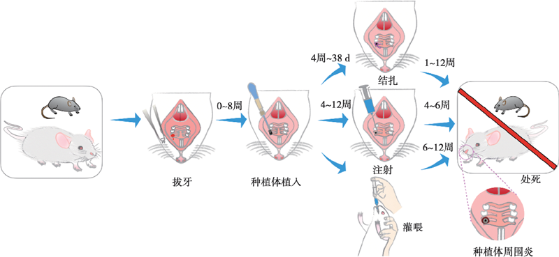
Fig.1
Flow chart of peri-implantitis model construction in rodents"
| [1] |
Schwarz F, Derks J, Monje A, et al. Peri-implantitis[J]. J Periodontol, 2018, 89(Suppl 1):S267-S290.
doi: 10.1002/JPER.16-0350 |
| [2] |
Moraschini V, daCPoubel LA, Ferreira VF, et al. Evaluation of survival and success rates of dental implants reported in longitudinal studies with a follow-up period of at least 10 years: A systematic review[J]. Int J Oral Maxillofac Surg, 2015, 44(3):377-388.
doi: 10.1016/j.ijom.2014.10.023 |
| [3] |
Lee CT, Huang YW, Zhu L, et al. Prevalences of peri-implantitis and peri-implant mucositis: Systematic review and meta-analysis[J]. J Dent, 2017, 62: 1-12.
doi: 10.1016/j.jdent.2017.04.011 |
| [4] |
Renvert S, Polyzois I. Treatment of pathologic peri-implant pockets[J]. Periodontol 2000, 2018, 76(1):180-190.
doi: 10.1111/prd.12149 |
| [5] |
Pearce AI, Richards RG, Milz S, et al. Animal models for implant biomaterial research in bone: A review[J]. Eur Cell Mater, 2007, 13: 1-10.
pmid: 17334975 |
| [6] |
Wang S, Liu Y, Fang D, et al. The miniature pig: A useful large animal model for dental and orofacial research[J]. Oral Dis, 2007, 13(6):530-537.
doi: 10.1111/j.1601-0825.2006.01337.x pmid: 17944668 |
| [7] |
Vlahović Z, Marković A, Lazić Z, et al. Histopathological comparative analysis of periimplant bone inflammatory response after dental implant insertion using flap and flapless surgical technique. An experimental study in pigs[J]. Clin Oral Implants Res, 2017, 28(9):1067-1073.
doi: 10.1111/clr.2017.28.issue-9 |
| [8] | Park SY, Kim KH, Rhee SH, et al. An immediate peri-implantitis induction model to study regenerative peri-implantitistreatments[J]. Clin Oral Implants Res, 2017, 28(1):36-42. |
| [9] |
Wancket LM. Animal models for evaluation of bone implants and devices: Comparative bone structure and common model uses[J]. Vet Pathol, 2015, 52(5):842-850.
doi: 10.1177/0300985815593124 pmid: 26163303 |
| [10] |
Sousa V, Mardas N, Spratt D, et al. Experimental models for contamination of titanium surfaces and disinfection protocols[J]. Clin Oral Implants Res, 2016, 27(10):1233-1242.
doi: 10.1111/clr.2016.27.issue-10 |
| [11] |
Stübinger S, Dard M. The rabbit as experimental model for research in implant dentistry and related tissue regeneration[J]. J Invest Surg, 2013, 26(5):266-282.
doi: 10.3109/08941939.2013.778922 pmid: 23617292 |
| [12] |
Kantarci A, Hasturk H, van Dyke TE. Animal models for periodontal regeneration and peri-implant responses[J]. Periodontol 2000, 2015, 68(1):66-82.
doi: 10.1111/prd.12052 |
| [13] |
Hiyari S, Naghibi A, Wong R, et al. Susceptibility of different mouse strains to peri-implantitis[J]. J Periodontal Res, 2018, 53(1):107-116.
doi: 10.1111/jre.12493 pmid: 29044525 |
| [14] | Shuto T, Wachi T, Shinohara Y, et al. Increase in receptor activator of nuclear factor κB ligand/osteoprotegerin ratio in peri-implant gingiva exposed to Porphyromonas gingivalis lipopolysaccharide[J]. JDentSci, 2016, 11(1):8-16. |
| [15] |
Yue G, Edani H, Sullivan A, et al. Is maxillary diastema an appropriate site for implantation in rats?[J]. Int J Implant Dent, 2020, 6(1):8.
doi: 10.1186/s40729-019-0203-5 pmid: 32100121 |
| [16] | 冷春涛, 李婷, 高晓蔚. 4种白细胞介素在种植体周围炎分期模型中的表达分析[J]. 上海口腔医学, 2020, 29(2):162-167. |
| [17] |
Wong RL, Hiyari S, Yaghsezian A, et al. Comparing the healing potential of late-stage periodontitis and peri-implantitis[J]. J Oral Implantol, 2017, 43(6):437-445.
doi: 10.1563/aaid-joi-D-17-00157 pmid: 29064761 |
| [18] |
Ding L, Zhang P, Wang X, et al. A doxycycline-treated hydroxyapatite implant surface attenuates the progression of peri-implantitis: A radiographic and histological study in mice[J]. Clin Implant Dent Relat Res, 2019, 21(1):154-159.
doi: 10.1111/cid.2019.21.issue-1 |
| [19] |
Wong RL, Hiyari S, Yaghsezian A, et al. Early intervention of peri-implantitis and periodontitis using a mouse model[J]. J Periodontol, 2018, 89(6):669-679.
doi: 10.1002/JPER.17-0541 pmid: 29520950 |
| [20] |
Li H, Wang YF, Zhang D, et al. Glycemic fluctuation exacerbates inflammation and bone loss and alters microbiota profile around implants in diabetic mice with experimental peri-implantitis[J]. Int J Implant Dent, 2021, 7(1):79.
doi: 10.1186/s40729-021-00360-9 pmid: 34401982 |
| [21] | Nguyen Vo TN, Hao J, Chou J, et al. Ligature induced peri-implantitis: Tissue destruction and inflammatory progression in a murine model[J]. Clin Oral Implants Res, 2017, 28(2):129-136. |
| [22] |
Hiyari S, Wong RL, Yaghsezian A, et al. Ligature-induced peri-implantitis and periodontitis in mice[J]. J Clin Periodontol, 2018, 45(1):89-99.
doi: 10.1111/jcpe.12817 pmid: 28921659 |
| [23] |
Pirih FQ, Hiyari S, Barroso ADV, et al. Ligature-induced peri-implantitis in mice[J]. J Periodontal Res, 2015, 50(4):519-524.
doi: 10.1111/jre.12234 pmid: 25244403 |
| [24] |
Li H, Chen ZY, Zhong XH, et al. Mangiferin alleviates experimental peri-implantitis via suppressing interleukin-6 production and Toll-like receptor 2 signaling pathway[J]. J Orthop Surg Res, 2019, 14(1):325.
doi: 10.1186/s13018-019-1387-3 |
| [25] |
Pan KQ, Hu Y, Wang YF, et al. RANKL blockade alleviates peri-implant bone loss and is enhanced by anti-inflammatory microRNA-146a through TLR2/4 signaling[J]. Int J Implant Dent, 2020, 6(1):15.
doi: 10.1186/s40729-020-00210-0 pmid: 32291538 |
| [26] |
Yu X, Hu Y, Freire M, et al. Role of toll-like receptor 2 in inflammation and alveolar bone loss in experimental peri-implantitis versus periodontitis[J]. J Periodontal Res, 2018, 53(1):98-106.
doi: 10.1111/jre.12492 pmid: 28872184 |
| [27] |
Yuan SS, Wang C, Jiang WT, et al. Comparative transcriptome analysis of gingival immune-mediated inflammation in peri-implantitis and periodontitis within the same host environment[J]. J Inflamm Res, 2022, 15:3119-3133.
doi: 10.2147/JIR.S363538 pmid: 35642216 |
| [28] |
Deng S, Hu Y, Zhou J, et al. TLR4 mediates alveolar bone resorption in experimental peri-implantitis through regulation of CD45+ cell infiltration, RANKL/OPG ratio, and inflammatory cytokine production[J]. J Periodontol, 2020, 91(5):671-682.
doi: 10.1002/JPER.18-0748 pmid: 31489644 |
| [29] |
Pirih FQ, Hiyari S, Leung HY, et al. A murine model of lipopolysaccharide-induced peri-implant mucositis and peri-implantitis[J]. J Oral Implantol, 2015, 41(5):e158-e164.
doi: 10.1563/aaid-joi-D-14-00068 |
| [30] |
Tzach-Nahman R, Mizraji G, Shapira L, et al. Oral infection with Porphyromonas gingivalis induces peri-implantitis in a murine model: Evaluation of bone loss and the local inflammatory response[J]. J Clin Periodontol, 2017, 44(7):739-748.
doi: 10.1111/jcpe.12735 pmid: 28453225 |
| [31] |
Varon-Shahar E, Shusterman A, Piattelli A, et al. Peri-implant alveolar bone resorption in an innovative peri-implantitis murine model: Effect of implant surface and onset of infection[J]. Clin Implant Dent Relat Res, 2019, 21(4):723-733.
doi: 10.1111/cid.v21.4 |
| [32] |
Hori Y, Kondo Y, Nodai T, et al. Xerostomia aggravates ligation-induced peri-implantitis: A preclinical in vivo study[J]. Clin Oral Implants Res, 2021, 32(5):581-589.
doi: 10.1111/clr.v32.5 |
| [33] |
Jung HJ, Lee W, Shin JS, et al. The effects of NF-kB inhibition with p65-TMD-linked PTD on inflammatory responses at peri-implantitissites[J]. Inflammation, 2021, 44(6):2291-2301.
doi: 10.1007/s10753-021-01500-4 |
| [34] |
Yamazaki S, Masaki C, Nodai T, et al. The effects of hyperglycaemia on peri-implant tissues after osseointegration[J]. J Prosthodont Res, 2020, 64(2):217-223.
doi: S1883-1958(19)30072-6 pmid: 31852608 |
| [35] | He Q, Mu Z, Shrestha A, et al. Development of a rat model for type 2 diabetes mellitus peri-implantitis: A preliminary study[J]. Oral Dis, 2021: 2021. |
| [36] |
Koutouzis T, Eastman C, Chukkapalli S, et al. A novel rat model of polymicrobialperi-implantitis: A preliminary study[J]. J Periodontol, 2017, 88(2):e32-e41.
doi: 10.1902/jop.2016.160273 |
| [37] |
Rovin S, Costich ER, Gordon HA. The influence of bacteria and irritation in the initiation of periodontal disease in germfree and conventional rats[J]. J Periodontal Res, 1966, 1(3):193-204.
pmid: 4225530 |
| [38] |
Wu XB, Qiao SC, Wang W, et al. Melatonin prevents peri-implantitis via suppression of TLR4/NF-κB[J]. Acta Biomater, 2021, 134: 325-336.
doi: 10.1016/j.actbio.2021.07.017 pmid: 34271168 |
| [39] |
Graves DT, Fine D, Teng YTA, et al. The use of rodent models to investigate host-bacteria interactions related to periodontal diseases[J]. J Clin Periodontol, 2008, 35(2):89-105.
doi: 10.1111/j.1600-051X.2007.01172.x pmid: 18199146 |
| [40] |
Lin J, Bi LJ, Yu XQ, et al. Porphyromonas gingivalis exacerbates ligature-induced, RANKL-dependent alveolar bone resorption via differential regulation of Toll-like receptor 2 (TLR2) and TLR4[J]. Infect Immun, 2014, 82(10):4127-4134.
doi: 10.1128/IAI.02084-14 |
| [41] |
Freire MO, Sedghizadeh PP, Schaudinn C, et al. Development of an animal model for Aggregatibacter actinomycetemcomitans biofilm-mediated oral osteolytic infection: A preliminary study[J]. J Periodontol, 2011, 82(5):778-789.
doi: 10.1902/jop.2010.100263 |
| [42] |
Becker ST, Föge M, Beck-Broichsitter BE, et al. Induction of periimplantitis in dental implants[J]. J Craniofac Surg, 2013, 24(1):e15-e18.
doi: 10.1097/SCS.0b013e318266fb2d |
| [43] |
李星佳, 陈琪欣, 袁长永, 等. 种植体周围炎大鼠模型研究[J]. 口腔医学研究, 2021, 37(4):314-318.
doi: 10.13701/j.cnki.kqyxyj.2021.04.009 |
| [44] | Sun JQ, Eberhard J, Glage S, et al. Development of a peri-implantitis model in the rat[J]. Clin Oral Implants Res, 2020, 31(3):203-214. |
| [45] |
Mori G, Sasaki H, Makabe Y, et al. The genes Scgb1a1, Lpo and Gbp2 characteristically expressed in peri-implant epithelium of rats[J]. Clin Oral Implants Res, 2016, 27(12):e190-e198.
doi: 10.1111/clr.2016.27.issue-12 |
| [46] |
Kotsakis GA, Olmedo DG. Peri-implantitis is not periodontitis: Scientific discoveries shed light on microbiome-biomaterial interactions that may determine disease phenotype[J]. Periodontol 2000, 2021, 86(1):231-240.
doi: 10.1111/prd.v86.1 |
| [47] | Takamori Y, Atsuta I, Nakamura H, et al. Histopathological comparison of the onset of peri-implantitis and periodontitis in rats[J]. Clin Oral Implants Res, 2017, 28(2):163-170. |
| [48] | Papi P, Letizia C, Pilloni A, et al. Peri-implant diseases and metabolic syndrome components: A systematic review[J]. Eur Rev Med Pharmacol Sci, 2018, 22(4):866-875. |
| [49] |
Washio K, Tsutsumi Y, Tsumanuma Y, et al. In vivo periodontium formation around titanium implants using periodontal ligament cell sheet[J]. Tissue Eng Part A, 2018, 24(15/16):1273-1282.
doi: 10.1089/ten.tea.2017.0405 |
| [50] |
Palmquist A, Johansson A, Suska F, et al. Acute inflammatory response to laser-induced micro- and nano-sized titanium surface features[J]. Clin Implant Dent Relat Res, 2013, 15(1):96-104.
doi: 10.1111/cid.2013.15.issue-1 |
| [51] |
Hasani-Sadrabadi MM, Sarrion P, Pouraghaei S, et al. An engineered cell-laden adhesive hydrogel promotes craniofacial bone tissue regeneration in rats[J]. Sci Transl Med, 2020, 12(534):eaay6853.
doi: 10.1126/scitranslmed.aay6853 |
| [52] |
Lee YH, Kim JS, Kim JE, et al. Nanoparticle mediated PPARγ gene delivery on dental implants improves osseointegration via mitochondrial biogenesis in diabetes mellitus rat model[J]. Nanomed Nanotechnol Biol Med, 2017, 13(5):1821-1832.
doi: 10.1016/j.nano.2017.02.020 |
| [53] |
Schwarz F, Sculean A, Engebretson SP, et al. Animal models for peri-implant mucositis and peri-implantitis[J]. Periodontol 2000, 2015, 68(1):168-181.
doi: 10.1111/prd.12064 |
| [54] |
Fine DH, Goncharoff P, Schreiner H, et al. Colonization and persistence of rough and smooth colony variants of Actinobacillusactinomycetemcomitans in the mouths of rats[J]. Arch Oral Biol, 2001, 46(11):1065-1078.
pmid: 11543714 |
| [55] |
Bai L, Chen BY, Liu Y, et al. A mouse periodontitis model with humanized oral bacterial community[J]. Front Cell Infect Microbiol, 2022, 12: 842845.
doi: 10.3389/fcimb.2022.842845 |
| Viewed | ||||||
|
Full text |
|
|||||
|
Abstract |
|
|||||