
Stomatology ›› 2023, Vol. 43 ›› Issue (11): 1041-1046.doi: 10.13591/j.cnki.kqyx.2023.11.014
• Summary • Previous Articles Next Articles
YE Hengni,CHEN Xuepeng,TANG Kuangyun,ZHOU Mengqi,HU Ji’an( )
)
Revised:2023-03-28
Online:2023-11-28
Published:2023-11-21
CLC Number:
YE Hengni, CHEN Xuepeng, TANG Kuangyun, ZHOU Mengqi, HU Ji’an. Progress of research on effect of Le Fort Ⅰ maxillary osteotomy on three-dimensional nasal morphology[J]. Stomatology, 2023, 43(11): 1041-1046.

Fig.1
Frontal view of the nose"
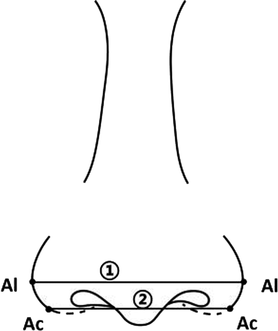
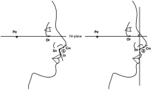
Fig.2
Profile of the nose"

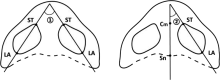
Fig.3
Bottom view of the nose"

| [1] |
Heiman AJ, Nair L, Kanth A, et al. Defining regional variation in nasal anatomy to guide ethnic rhinoplasty:A systematic review[J]. J Plast Reconstr Aesthetic Surg, 2022, 75(8):2784-2795.
doi: 10.1016/j.bjps.2022.04.058 |
| [2] |
Worasakwutiphong S, Chuang YF, Chang HW, et al. Nasal changes after orthognathic surgery for patients with prognathism and Class Ⅲ malocclusion:Analysis using three-dimensional photogrammetry[J]. J Formos Med Assoc, 2015, 114(2):112-123.
doi: 10.1016/j.jfma.2014.10.003 |
| [3] |
Hellak AF, Kirsten B, Schauseil M, et al. Influence of maxillary advancement surgery on skeletal and soft-tissue changes in the nose—a retrospective cone-beam computed tomography study[J]. Head Face Med, 2015, 11(1):23.
doi: 10.1186/s13005-015-0080-y |
| [4] |
Allar ML, Movahed R, Wolford LM, et al. Nasolabial changes following double jaw surgery[J]. J Craniofac Surg, 2019, 30(8):2560-2564.
doi: 10.1097/SCS.0000000000005876 pmid: 31689731 |
| [5] |
Rohrich RJ, Malafa MM, Ahmad J, et al. Managing alar flare in rhinoplasty[J]. Plast Reconstr Surg, 2017, 140(5):910-919.
doi: 10.1097/PRS.0000000000003786 pmid: 29068925 |
| [6] |
Moon KC, Han SK. Surgical anatomy of the Asian nose[J]. Facial Plast Surg Clin North Am, 2018, 26(3):259-268.
doi: 10.1016/j.fsc.2018.03.001 |
| [7] |
Choi SY, Kim SJ, Lee HY, et al. Esthetic nasolabial angle according to the degree of upper lip protrusion in an Asian population[J]. Am J Rhinol Allergy, 2018, 32(1):66-70.
doi: 10.2500/ajra.2018.32.4485 pmid: 29336294 |
| [8] |
Ahmed O, Dhinsa A, Popenko N, et al. Population-based assessment of currently proposed ideals of nasal tip projection and rotation in young women[J]. JAMA Facial Plast Surg, 2014, 16(5):310-318.
doi: 10.1001/jamafacial.2014.228 |
| [9] |
Ganske IM, Tan RA, Langa OC, et al. Does the nostril shape change after le fort I advancement in patients with unilateral complete cleft lip?[J]. J Oral Maxillofac Surg, 2020, 78(6):998-1005.
doi: 10.1016/j.joms.2020.01.010 |
| [10] | Suzen M, Dilaver E, Ak KB, et al. Analysis of inferior nasal morphology and nostrils following le fort I osteotomy[J]. J CraniofacSurg, 2022, 33(8):2682-2687. |
| [11] | Dindaroğlu F, Kutlu P, Duran GS, et al. Accuracy and reliability of 3D stereophotogrammetry:A comparison to direct anthropometry and 2D photogrammetry[J]. Angle Orthod, 2016, 86(3):487-494. |
| [12] | Atakan A, Özçırpıcı AA. Correlation between cephalometric nasal changes and patients’ perception after orthognathic surgery[J]. Am J Orthod Dentofac Orthop, 2021, 159(6):e449-e460. |
| [13] |
Baysal A, Sahan AO, Ozturk MA, et al. Reproducibility and reliability of three-dimensional soft tissue landmark identification using three-dimensional stereophotogrammetry[J]. Angle Orthod, 2016, 86(6):1004-1009.
pmid: 27023408 |
| [14] |
Guntaka PK, Kiang K, Caprio R, et al. Do patients treated with Invisalign have less swelling after orthognathic surgery than those with fixed orthodontic appliances?[J]. Am J Orthod Dentofac Orthop, 2023, 163(2):243-251.
doi: 10.1016/j.ajodo.2021.11.015 |
| [15] |
Coban G, Yavuz I, Karadas B, et al. Three-dimensional assess-ment of nasal changes after maxillary advancement with impaction using stereophotogrammetry[J]. Korean J Orthod, 2020, 50(4):249-257.
doi: 10.4041/kjod.2020.50.4.249 |
| [16] |
Almukhtar A, Khambay B, Ju X, et al. Comprehensive analysis of soft tissue changes in response to orthognathic surgery:Mandibular versus bimaxillary advancement[J]. Int J Oral Maxillofac Surg, 2018, 47(6):732-737.
doi: 10.1016/j.ijom.2017.11.014 |
| [17] |
Jung J, Lee CH, Lee JW, et al. Three dimensional evaluation of soft tissue after orthognathic surgery[J]. Head Face Med, 2018, 14(1):21.
doi: 10.1186/s13005-018-0179-z pmid: 30290762 |
| [18] |
D’Ettorre G, Farronato M, Candida E, et al. A comparison between stereophotogrammetry and smartphone structured light technology for three-dimensional face scanning[J]. Angle Orthod, 2022, 92(3):358-363.
doi: 10.2319/040921-290.1 pmid: 35015071 |
| [19] |
Pan FW, Liu JL, Cen YY, et al. Accuracy of RGB-D camera-based and stereophotogrammetric facial scanners:A comparative study[J]. J Dent, 2022, 127:104302.
doi: 10.1016/j.jdent.2022.104302 |
| [20] | Zhao YJ, Xiong YX, Wang Y. Three-dimensional accuracy of facial scan for facial deformities in clinics:A new evaluation method for facial scanner accuracy[J]. PLoS One, 2017, 12(1):e0169402. |
| [21] |
ten Harkel TC, Vinayahalingam S, Ingels KJAO, et al. Reliability and agreement of 3D anthropometric measurements in facial palsy patients using a low-cost 4D imaging system[J]. IEEE Trans Neural Syst Rehabil Eng, 2020, 28(8):1817-1824.
doi: 10.1109/TNSRE.7333 |
| [22] |
Petrides G, Clark JR, Low H, et al. Three-dimensional scanners for soft-tissue facial assessment in clinical practice[J]. J Plast Reconstr Aesthetic Surg, 2021, 74(3):605-614.
doi: 10.1016/j.bjps.2020.08.050 |
| [23] |
Chen ZC, Albdour MN, Lizardo JA, et al. Precision of three-dimensional stereo-photogrammetry (3dMDTM) in anthropometry of the auricle and its application in microtiare construction[J]. J Plast Reconstr Aesthetic Surg, 2015, 68(5):622-631.
doi: 10.1016/j.bjps.2015.02.020 |
| [24] |
Raffone C, Gianfreda F, Pompeo MG, et al. Chairside virtual patient protocol. Part 2:Management of multiple face scans and alignment predictability[J]. J Dent, 2022, 122:104123.
doi: 10.1016/j.jdent.2022.104123 |
| [25] |
Thurzo A, Strunga M, Havlínová R, et al. Smartphone-based facial scanning as a viable tool for facially driven orthodontics?[J]. Sensors (Basel), 2022, 22(20):7752.
doi: 10.3390/s22207752 |
| [26] |
Liu JL, Zhang CH, Cai RL, et al. Accuracy of 3-dimensional stereophotogrammetry: Comparison of the 3dMD and Bellus3D facial scanning systems with one another and with direct anthropometry[J]. Am J Orthod Dentofacial Orthop, 2021, 160(6):862-871.
doi: 10.1016/j.ajodo.2021.04.020 |
| [27] |
Seon S, Lee HW, Jeong BJ, et al. Study of soft tissue changes in the upper lip and nose after backward movement of the maxilla in orthognathic surgery[J]. J Korean Assoc Oral Maxillofac Surg, 2020, 46(6):385-392.
doi: 10.5125/jkaoms.2020.46.6.385 pmid: 33377463 |
| [28] |
Lee JY, Kim YI, Hwang DS, et al. Effect of setback Le Fort I osteotomy on midfacial soft-tissue changes as evaluated by cone-beam computed tomography superim position for cases of skeletal Class Ⅲ malocclusion[J]. Int J Oral Maxillofac Surg, 2013, 42(6):790-795.
doi: 10.1016/j.ijom.2012.11.012 |
| [29] | Sabri H, Tehranchi A, Sarkarat F. 3-dimensional analysis of nasal soft tissue alterations following maxillary Lefort I advancement with and without impaction using 3D photogrammetry scanner[J]. Oral Maxillofac Surg, 2022:1-13. |
| [30] |
Denadai R, Chou PY, Yao CF, et al. Effect of le fort I maxillary repositioning on three-dimensional nasal tip rotation:A comparative study with implication for the Asian nose[J]. Plast Reconstr Surg, 2021, 147(4):903-914.
doi: 10.1097/PRS.0000000000007774 pmid: 33750094 |
| [31] |
Denadai R, Chou PY, Lin YY, et al. Type of maxillary segment mobilization affects three-dimensional nasal morphology[J]. J Plast Reconstr Aesthetic Surg, 2021, 74(3):592-604.
doi: 10.1016/j.bjps.2020.08.119 |
| [32] |
de Sousa Gil AP, Guijarro-Martínez R, Haas OL, et al. Three-dimensional analysis of nasolabial soft tissue changes after Le Fort I osteotomy:A systematic review of the literature[J]. Int J Oral Maxillofac Surg, 2019, 48(9):1185-1200.
doi: 10.1016/j.ijom.2019.01.028 |
| [33] |
Üstün GG, Konaş E, El H, et al. The effects of maxillary movements on nasal aesthetics following orthognathic surgery[J]. J Craniofac Surg, 2020, 31(3):796-800.
doi: 10.1097/SCS.0000000000006167 pmid: 31934978 |
| [34] |
Sawh-Martinez R, Lin AM, DeSesa CR, et al. Clockwise and counterclockwise le fort I movements influence nasolabial morphology differently[J]. Plast Reconstr Surg, 2018, 142(6):1572-1581.
doi: 10.1097/PRS.0000000000004988 pmid: 30188468 |
| [35] |
Schendel SA, Williamson LW. Muscle reorientation following superior repositioning of the maxilla[J]. J Oral Maxillofac Surg, 1983, 41(4):235.
doi: 10.1016/0278-2391(83)90265-3 |
| [36] |
Khamashta-Ledezma L, Naini FB. Prospective assessment of maxillary advancement effects:Maxillary incisor exposure, and upper lip and nasal changes[J]. Am J Orthod Dentofac Orthop, 2015, 147(4):454-464.
doi: 10.1016/j.ajodo.2014.11.028 |
| [37] |
Westermark AH, Bystedt H, von Konow L, et al. Nasolabial morphology after le fort I osteotomies effect of alar base suture[J]. Int J Oral Maxillofac Surg, 1991, 20(1):25-30.
doi: 10.1016/S0901-5027(05)80690-3 |
| [38] |
Muradin MSM, Rosenberg A, van der Bilt A, et al. The effect of alar cinch sutures and V-Y closure on soft tissue dynamics after Le Fort I intrusion osteotomies[J]. J Cranio Maxillofac Surg, 2009, 37(6):334-340.
doi: 10.1016/j.jcms.2009.03.004 |
| [39] | Monnazzi MS, Mannarino FS, Gabrielli MFR. Extraoral alar base cinch. A modification for the technique[J]. J Oral Maxillofac Surg Med Pathol, 2014, 26(2):142-144. |
| [40] |
Mani V, Panicker P, Shenoy A, et al. Evaluation of changes in the alar base width following lefort 1 and AMO with conventional alar cinch suturing:A photographic study of 100 cases[J]. J Maxillofac Oral Surg, 2020, 19(1):21-25.
doi: 10.1007/s12663-019-01227-8 |
| [41] |
Raithatha R, Naini FB, Patel S, et al. Long-term stability of limiting nasal alar base width changes with a cinch suture following Le Fort I osteotomy with submentalintubation[J]. Int J Oral Maxillofac Surg, 2017, 46(11):1372-1379.
doi: 10.1016/j.ijom.2017.04.027 |
| [42] |
Mahsoub R, Naini FB, Patel S, et al. Nasolabial angle and nasal tip elevation changes in profile view following a Le Fort I osteotomy with or without the use of an alar base cinch suture:A long-term cohort study[J]. Oral Surg Oral Med Oral Pathol Oral Radiol, 2020, 130(4):379-386.
doi: 10.1016/j.oooo.2020.05.011 |
| [43] |
Ishida T, Manabe A, Yang SS, et al. An orthodontic-orthognathic patient with obstructive sleep apnea treated with Le Fort I osteotomy advancement and alar cinch suture combined with a muco-musculo-periosteal V-Y closure to minimize nose deformity[J]. Angle Orthod, 2019, 89(6):946-952.
doi: 10.2319/052818-406.1 pmid: 30698453 |
| [44] | Yang J, Chu Y, Liao H, et al. Comparison of the effectiveness between conventional and modified cinch suture techniques in LeFort I osteotomy:A systematic review and meta-analysis of randomized control trials[J]. Ann Plast Surge, 2023,doi:10.1097/SAP.0000000000003354. |
| [45] |
Gil APS, Machado-Fernández A, Guijarro-Martínez R, et al. Le Fort I osteotomy and soft tissue response:A retrospective cohort study comparing three different techniques[J]. J Cranio Maxillofac Surg, 2022, 50(2):107-113.
doi: 10.1016/j.jcms.2021.11.009 |
| [46] |
Cho YS, Hwang KG, Park CJ. Postoperative effects of anterior nasal spine bone harvesting on overall nasal shape[J]. Clin Oral Implants Res, 2013, 24(6):618-622.
doi: 10.1111/clr.2013.24.issue-6 |
| [47] |
Jung S, Kim JY, Jung YS, et al. Reliability of anterior nasal spine as a reference point after LeFort I surgery using three-dimensional analysis[J]. J Craniofac Surg, 2022, 33(7):2104-2108.
doi: 10.1097/SCS.0000000000008619 pmid: 35261362 |
| [48] |
Zhong YH, Zhu YJ, Jiang TR, et al. A novel study on alar mobility of HAN female by 3dMD dynamic surface imaging system[J]. Aesth Plast Surg, 2022, 46(1):364-372.
doi: 10.1007/s00266-021-02386-1 |
| Viewed | ||||||||||||||||||||||||||||||||||||||||||||||||||
|
Full text 408
|
|
|||||||||||||||||||||||||||||||||||||||||||||||||
|
Abstract 414
|
|
|||||||||||||||||||||||||||||||||||||||||||||||||