
Stomatology ›› 2023, Vol. 43 ›› Issue (10): 865-871.doi: 10.13591/j.cnki.kqyx.2023.10.001
• Basic Research • Next Articles
LIU Yang,LI Qiang,LEI Rong,CHEN Yongjin,ZHAO Yajuan( )
)
Revised:2023-06-27
Online:2023-10-28
Published:2023-10-20
CLC Number:
LIU Yang, LI Qiang, LEI Rong, CHEN Yongjin, ZHAO Yajuan. The involvement of trigeminal motor nucleus in the electromyography change of masseter muscle induced by chronic restraint stress in mice[J]. Stomatology, 2023, 43(10): 865-871.
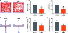
Fig.1
Behavioral changes in the OF and EPM after chronic restraint stress"
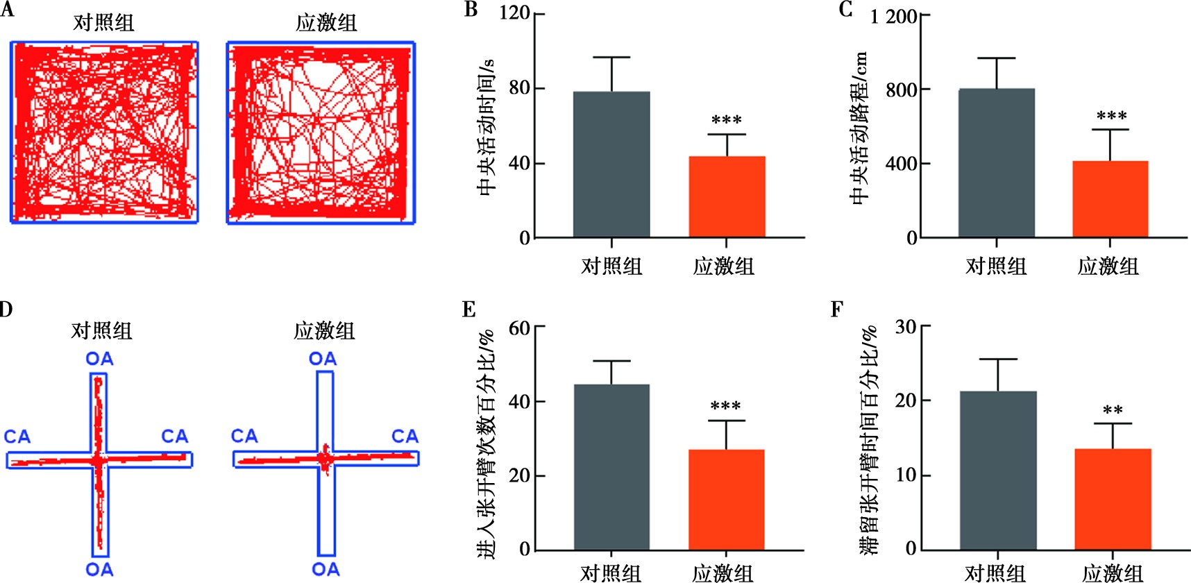
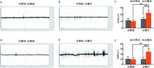
Fig.2
The EMG level of masseter muscle before and after chronic restraint stress"
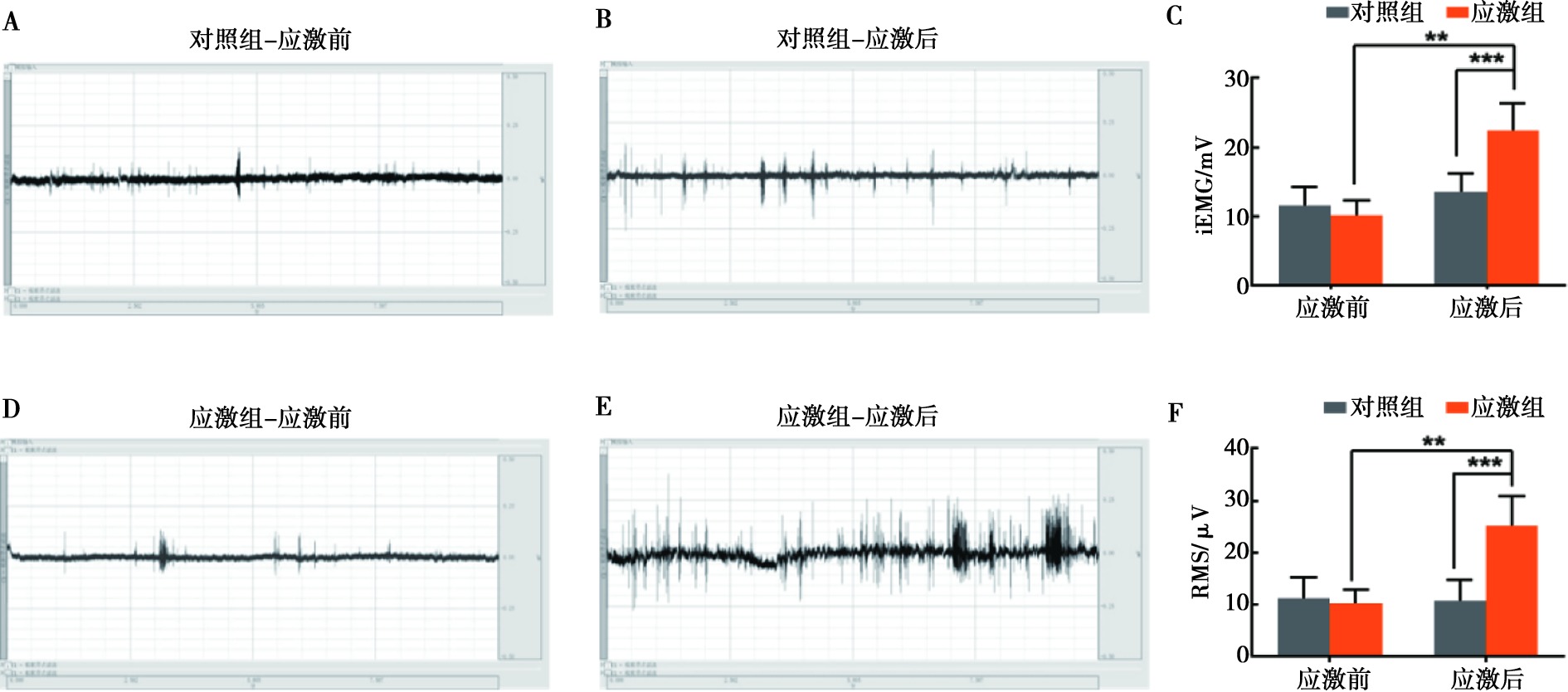
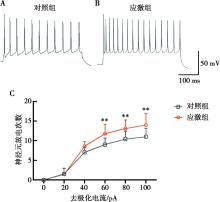
Fig.3
The spike numbers of Vmo neurons after chronic restraint stress"
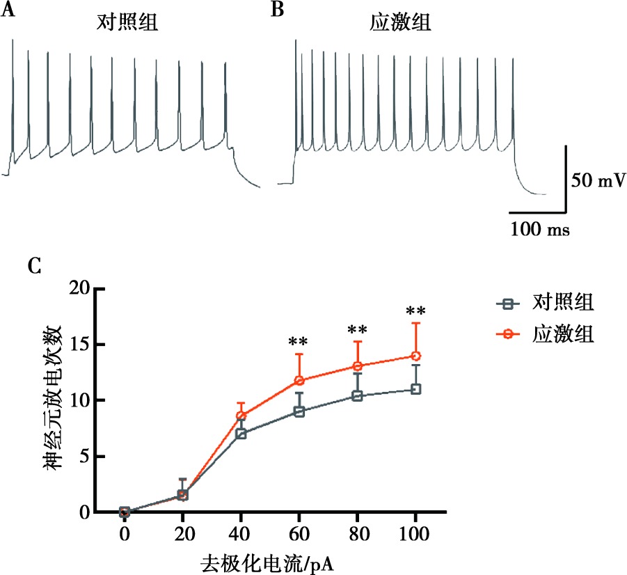

Fig.4
The sEPSCs of Vmo neurons after chronic restraint stress"
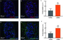
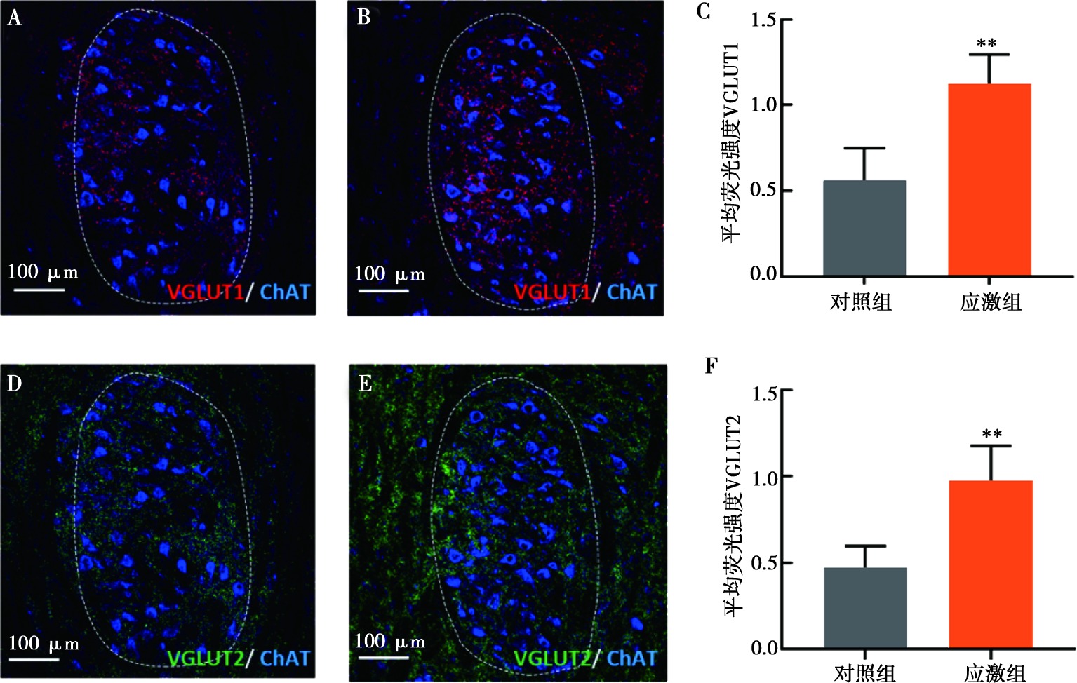
Fig.5
Immunofluorescence staining results of VGLUT1 and VGLUT2 in Vmo neurons after chronic restraint stress"
| [1] | 傅开元. 2014年新版国际颞下颌关节紊乱病分类及诊断标准解读[J]. 中华口腔医学杂志, 2017, 52(6):374-376. |
| [2] |
Slade GD, Ohrbach R, Greenspan JD, et al. Painful temporomandibular disorder:Decade of discovery from OPPERA studies[J]. J Dent Res, 2016, 95(10):1084-1092.
doi: 10.1177/0022034516653743 pmid: 27339423 |
| [3] | 徐文鑫, 马宇锋. 声乐生颞下颌关节紊乱病问卷调查及危险因素分析[J]. 口腔医学, 2023, 43(4):343-346. |
| [4] |
Yap AU, Zhang MJ, Cao Y, et al. Comparison of psychological states and oral health-related quality of life of patients with differing severity of temporomandibular disorders[J]. J Oral Rehabil, 2022, 49(2):177-185.
doi: 10.1111/joor.v49.2 |
| [5] |
Zieliński G, Ginszt M, Zawadka M, et al. The relationship between stress and masticatory muscle activity in female students[J]. J Clin Med, 2021, 10(16):3459.
doi: 10.3390/jcm10163459 |
| [6] |
Janal MN, Lobbezoo F, Quigley KS, et al. Stress-evoked muscle activity in women with and without chronic myofascial face pain[J]. J Oral Rehabil, 2021, 48(10):1089-1098.
doi: 10.1111/joor.13238 pmid: 34370315 |
| [7] |
Kitagawa K, Kodama N, Manda Y, et al. Effect of masseter muscle activity during wakefulness and sleep on tooth wear[J]. J Prosthodont Res, 2021, 66(4):551-556.
doi: 10.2186/jpr.JPR_D_21_00171 pmid: 34955483 |
| [8] |
Choi KH, Kwon OS, Kim L, et al. Electromyographic changes in masseter and sternocleidomastoid muscles can be applied to diagnose of temporomandibular disorders:An observational study[J]. Integr Med Res, 2021, 10(4):100732.
doi: 10.1016/j.imr.2021.100732 |
| [9] |
Dinsdale A, Liang ZQ, Thomas L, et al. Is jaw muscle activity impaired in adults with persistent temporomandibular disorders? A systematic review and meta-analysis[J]. J Oral Rehabil, 2021, 48(4):487-516.
doi: 10.1111/joor.13139 pmid: 33369753 |
| [10] |
Slaoui Hasnaoui M, Arsenault I, Verdier D, et al. Functional connectivity between the trigeminal main sensory nucleus and the trigeminal motor nucleus[J]. Front Cell Neurosci, 2020, 14:167.
doi: 10.3389/fncel.2020.00167 pmid: 32655373 |
| [11] |
Faunes M, Oñate-Ponce A, Fernández-Collemann S, et al. Excitatory and inhibitory innervation of the mouse orofacial motor nuclei:A stereological study[J]. J Comp Neurol, 2016, 524(4):738-758.
doi: 10.1002/cne.v524.4 |
| [12] |
Zhang FX, Ge SN, Dong YL, et al. Vesicular glutamate transpo-rter isoforms:The essential players in the somatosensory systems[J]. Prog Neurobiol, 2018, 171:72-89.
doi: 10.1016/j.pneurobio.2018.09.006 |
| [13] |
Park SK, Ko SJ, Paik SK, et al. Vesicular glutamate transporter 1 (VGLUT1)- and VGLUT2-immunopositive axon terminals on the rat jaw-closing and jaw-opening motoneurons[J]. Brain Struct Funct, 2018, 223(5):2323-2334.
doi: 10.1007/s00429-018-1636-y pmid: 29476240 |
| [14] |
Abe C, Inoue T, Inglis MA, et al. C1 neurons mediate a stress-induced anti-inflammatory reflex in mice[J]. Nat Neurosci, 2017, 20(5):700-707.
doi: 10.1038/nn.4526 pmid: 28288124 |
| [15] | Segklia K, Stamatakis A, Stylianopoulou F, et al. Increased anxiety-related behavior, impaired cognitive function and cellular alterations in the brain of Cend1-deficient mice[J]. Front Cell Neurosci, 2019, 12:497. |
| [16] |
Zhao YJ, Liu Y, Wang J, et al. Activation of the mesencephalic trigeminal nucleus contributes to masseter hyperactivity induced by chronic restraint stress[J]. Front Cell Neurosci, 2022, 16:841133.
doi: 10.3389/fncel.2022.841133 |
| [17] |
Yu H, Xiang XK, Chen ZM, et al. Periaqueductal gray neurons encode the sequential motor program in hunting behavior of mice[J]. Nat Commun, 2021, 12(1):6523.
doi: 10.1038/s41467-021-26852-1 pmid: 34764279 |
| [18] | Paxinos G, Franklin KBJ. The mouse brain in stereotaxic coordinates[M]. Cambridge: Academic Press, 2001. |
| [19] |
Atefeh H, Nazanin M, Berdi ODR, et al. Personality traits and anxiety in patients with temporomandibular disorders[J]. BMC Psychol, 2022, 10(1):86.
doi: 10.1186/s40359-022-00795-8 pmid: 35379356 |
| [20] |
Huo R, Zeng BH, Zeng L, et al. Microbiota modulate anxiety-like behavior and endocrine abnormalities in hypothalamic-pituitary-adrenal axis[J]. Front Cell Infect Microbiol, 2017, 7:489.
doi: 10.3389/fcimb.2017.00489 |
| [21] |
Liu WZ, Zhang WH, Zheng ZH, et al. Identification of a prefrontal cortex-to-amygdala pathway for chronic stress-induced anxiety[J]. Nat Commun, 2020, 11(1):2221.
doi: 10.1038/s41467-020-15920-7 |
| [22] |
Shah N, Melo L, Reid WD, et al. Masseter deoxygenation in adults at risk for temporomandibular disorders[J]. J Dent Res, 2019, 98(6):666-672.
doi: 10.1177/0022034519837249 pmid: 30946624 |
| [23] |
Miao L, Huang F, Sun YY, et al. Curcumin plays a local anti-inflammatory and antioxidant role via the HMGB1/TLR4/NF-κB pathway in rat masseter muscle under psychological stress[J]. J Oral Rehabil, 2022, 49(2):249-257.
doi: 10.1111/joor.v49.2 |
| [24] |
Lin WQ, Zhao YJ, Cheng BX, et al. NMDAR and JNK activation in the spinal trigeminal nucleus caudalis contributes to masseter hyperalgesia induced by stress[J]. Front Cell Neurosci, 2019, 13:495.
doi: 10.3389/fncel.2019.00495 pmid: 31798413 |
| [25] |
Nakamura S, Nakayama K, Mochizuki A, et al. Electrophysiological and morphological properties of rat supratrigeminal premotor neurons targeting the trigeminal motor nucleus[J]. J Neurophysiol, 2014, 111(9):1770-1782.
doi: 10.1152/jn.00276.2013 pmid: 24501266 |
| [26] |
Han WF, Tellez LA, Rangel MJ, et al. Integrated control of predatory hunting by the central nucleus of the amygdala[J]. Cell, 2017, 168(1/2):311-324.e18.
doi: 10.1016/j.cell.2016.12.027 |
| [27] |
Mascaro MB, Prosdócimi FC, Bittencourt JC, et al. Forebrain projections to brainstem nuclei involved in the control of mandibular movements in rats[J]. Eur J Oral Sci, 2009, 117(6):676-684.
doi: 10.1111/j.1600-0722.2009.00686.x pmid: 20121930 |
| [28] |
Kaya BT, Geha P, de Araujo I, et al. Identification of central amygdala and trigeminal motor nucleus connectivity in humans:An ultra-high field diffusion MRI study[J]. Hum Brain Mapp, 2023, 44(4):1309-1319.
doi: 10.1002/hbm.v44.4 |
| Viewed | ||||||
|
Full text |
|
|||||
|
Abstract |
|
|||||