
Stomatology ›› 2023, Vol. 43 ›› Issue (10): 930-934.doi: 10.13591/j.cnki.kqyx.2023.10.013
• Summary • Previous Articles Next Articles
QIAN Min,WU Mengjie( )
)
Revised:2023-03-10
Online:2023-10-28
Published:2023-10-20
CLC Number:
QIAN Min, WU Mengjie. Review of MRI-based three-dimensional model reconstruction of temporomandibular joint[J]. Stomatology, 2023, 43(10): 930-934.
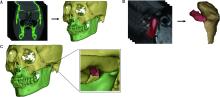
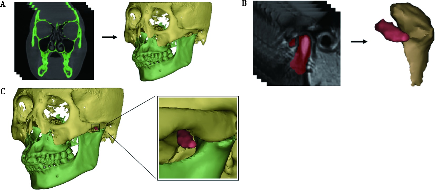
Fig.1
Three-dimensional reconstruction and registration of temporomandibular joint"
| [1] |
Somay E, Yilmaz B. Comparison of clinical and magnetic resona-nce imagining data of patients with temporomandibular disorders[J]. Niger J Clin Pract, 2020, 23(3):376-380.
doi: 10.4103/njcp.njcp_492_19 pmid: 32134038 |
| [2] | 张利, 李生娇, 施雄, 等. 口腔全景片图像质量影响因素分析[J]. 口腔颌面外科杂志, 2018, 28(4):225-228. |
| [3] | 李源, 刘木清, 刘博, 等. 基于锥形束CT影像的颞下颌关节髁突骨改建三维定量评价研究[J]. 中华口腔医学杂志, 2020, 55(9):617-623. |
| [4] | 何冬梅, 张露珠, 杨驰. 国产人工颞下颌关节的研发和初步临床应用[J]. 中华口腔医学杂志, 2017, 52(4):232-237. |
| [5] | 王文晖, 甄俊平. 颞下颌关节紊乱病MRI研究进展[J]. 磁共振成像, 2022, 13(5):148-150, 166. |
| [6] | 高辉, 邱兆文. 三维重建技术在临床医学中的应用[J]. 软件导刊, 2018, 17(6):170-172, 177. |
| [7] | 王昊, 康晓东, 刘玲玲, 等. MR图像去噪算法研究进展[J]. 磁共振成像, 2012, 3(3):231-236. |
| [8] | Rao GS, Srinivas B. De-noising of MRI brain tumor image using deep convolutional neural network[J]. SSRN J, 2019, 26:1192-1197. |
| [9] |
Jiang DS, Dou WQ, Vosters L, et al. Denoising of 3D magnetic resonance images with multi-channel residual learning of convolutional neural network[J]. Jpn J Radiol, 2018, 36(9):566-574.
doi: 10.1007/s11604-018-0758-8 pmid: 29982919 |
| [10] |
Lurie A, Tosoni GM, Tsimikas J, et al. Recursive hierarchic segmentation analysis of bone mineral density changes on digital panoramic images[J]. Oral Surg Oral Med Oral Pathol Oral Radiol, 2012, 113(4):549-558.e1.
doi: 10.1016/j.oooo.2011.10.002 |
| [11] | Trivedi DN, Kothari AM, Shah S, et al. Dental image matching by canny algorithm for human identification[J]. Int J Adv Comput Res, 2014, 4(17):985-990. |
| [12] | Tikhe SV, Naik AM, Bhide SD, et al. Algorithm to identify enamel caries and interproximal caries using dental digital radiographs[C]// 2016 IEEE 6th International Conference on Advanced Computing (IACC). Bhimavaram, India. IEEE, 2016:225-228. |
| [13] |
Ito S, Mine Y, Yoshimi Y, et al. Automated segmentation of articular disc of the temporomandibular joint on magnetic resonance images using deep learning[J]. Sci Rep, 2022, 12(1):221.
doi: 10.1038/s41598-021-04354-w pmid: 34997167 |
| [14] |
Li MX, Punithakumar K, Major PW, et al. Temporomandibular joint segmentation in MRI images using deep learning[J]. J Dent, 2022, 127:104345.
doi: 10.1016/j.jdent.2022.104345 |
| [15] |
Nozawa M, Ito H, Ariji Y, et al. Automatic segmentation of the temporomandibular joint disc on magnetic resonance images using a deep learning technique[J]. Dentomaxillofac Radiol, 2022, 51(1):20210185.
doi: 10.1259/dmfr.20210185 |
| [16] |
Kao ZK, Chiu NT, Wu HT H, et al. Classifying temporomandibular disorder with artificial intelligent architecture using magnetic resonance imaging[J]. Ann Biomed Eng, 2023, 51(3):517-526.
doi: 10.1007/s10439-022-03056-2 |
| [17] | Chen ZL, Xu JL, Shen YQ, et al. Application of CT medical imaging combined with deep learning 3D reconstruction in the diagnosis and rehabilitation of anterior cruciate ligament injury in table tennis players[J]. J HealthcEng, 2021, 2021:1152368. |
| [18] |
Khan U, Yasin A, Abid M, et al. A methodological review of 3D reconstruction techniques in tomographic imaging[J]. J Med Syst, 2018, 42(10):190.
doi: 10.1007/s10916-018-1042-2 pmid: 30178184 |
| [19] | 方威扬, 林东鑫, 寇万福, 等. 医学图像三维重建系统的研究进展[J]. 中国医学物理学杂志, 2022, 39(7):823-827. |
| [20] | 艾松涛, 唐为卿, 戴尅戎, 等. 颞下颌关节三维动态磁共振成像研究及生物力学分析[J]. 医用生物力学, 2013, 28(1):79-84, 90. |
| [21] |
Shu JH, Teng HD, Shao BM, et al. Biomechanical responses of temporomandibular joints during the lateral protrusions:A 3D finite element study[J]. Comput Methods Programs Biomed, 2020, 195:105671.
doi: 10.1016/j.cmpb.2020.105671 |
| [22] |
Wang RY, Bi RY, Liu Y, et al. Morphological changes of TMJ disc in surgically treated ADDwoR patients:A retrospective study[J]. BMC Oral Health, 2022, 22(1):432.
doi: 10.1186/s12903-022-02469-8 |
| [23] | 赖林锋, 方一鸣, 吴立军, 等. 不可复性颞下颌关节盘前移位术前与术后可视性个体化建模[J]. 浙江创伤外科, 2010, 15(2):129-132. |
| [24] | 周子凌, 张渊, 石利强, 等. 咬合接触对颞下颌关节应力分布影响的三维有限元分析[J]. 中华口腔医学杂志, 2015, 50(5):302-306. |
| [25] | 周伟, 安金刚, 荣起国, 等. 下颌骨颏部骨折联合双侧髁突囊内骨折致伤机制的三维有限元分析[J]. 北京大学学报(医学版), 2021, 53(5):983-989. |
| [26] | 何妍明, 王荷燕, 冯亚平, 等. 颞下颌关节盘MRI和锥形束CT图像配准的初步研究[J]. 中华口腔医学杂志, 2020, 55(10):772-777. |
| [27] | 傅开元, 胡敏, 余强, 等. 颞下颌关节常规MRI检查规范及关节盘移位诊断标准的专家共识[J]. 中华口腔医学杂志, 2020, 55(9):608-612. |
| [28] | 陈立奇, 薛卓维, 吴氢凯. 基于磁共振成像的女性盆底器官三维数字模型重建的研究进展[J]. 上海交通大学学报(医学版), 2022, 42(3):381-386. |
| [29] |
Hayakawa Y, Kober C, Otonari-Yamamoto M, et al. An approach for three-dimensional visualization using high-resolution MRI of the temporomandibular joint[J]. Dentomaxillofac Radiol, 2007, 36(6):341-347.
doi: 10.1259/dmfr/12894471 |
| [30] |
Chirani RA, Jacq JJ, Meriot P, et al. Temporomandibular joint:A methodology of magnetic resonance imaging 3-D reconstruction[J]. Oral Surg Oral Med Oral Pathol Oral Radiol Endod, 2004, 97(6):756-761.
doi: 10.1016/j.tripleo.2004.02.073 |
| [31] | Coombs MC, Bonthius DJ, Nie XJ, et al. Effect of measurement technique on TMJ mandibular condyle and articular disc morphometry:CBCT, MRI, and physical measurements[J]. J Oral MaxillofacSurg, 2019, 77(1):42-53. |
| [32] |
Kinzinger GSM, Hourfar J, Kober C, et al. Mandibular fossa morphology during therapy with a fixed functional orthodontic appliance[J]. J Orofac Orthop / Fortschritte Der Kieferorthopädie, 2018, 79(2):116-132.
doi: 10.1007/s00056-018-0124-6 |
| [33] |
Yushkevich PA, Pashchinskiy A, Oguz I, et al. User-guided segmentation of multi-modality medical imaging datasets with ITK-SNAP[J]. Neuroinformatics, 2019, 17(1):83-102.
doi: 10.1007/s12021-018-9385-x pmid: 29946897 |
| [34] | Luo D, Yang Z, Qiu C, et al. A magnetic resonance imaging study on the temporomandibular joint disc-condyle relationship in young asymptomatic adults[J]. Int J Oral Maxillofac Surg, 2022, 51(2):226-233. |
| [35] | NascimentoFalcão I, Cal Alonso MBC, da Silva LH, et al. 3D morphology analysis of TMJ articular eminence in magnetic resonance imaging[J]. Int J Dent, 2017, 2017:5130241. |
| [36] |
Costa ALF, Yasuda CL, Appenzeller S, et al. Comparison of conventional MRI and 3D reconstruction model for evaluation of temporomandibular joint[J]. Surg Radiol Anat, 2008, 30(8):663-667.
doi: 10.1007/s00276-008-0400-z pmid: 18704257 |
| [37] | Cookson JDE, Holman J. The illusion of solidity[J]. New Scientist, 1987, 6:50-53. |
| [38] |
Chu SA, Skultety KJ, Suvinen TI, et al. Computerized three-dimensional magnetic resonance imaging reconstructions of temporomandibular joints for both a model and patients with temporomandibular pain dysfunction[J]. Oral Surg Oral Med Oral Pathol Oral Radiol Endod, 1995, 80(5):604-611.
doi: 10.1016/S1079-2104(05)80157-X |
| [39] |
Chu SA, Suvinen TI, Clement JG, et al. The effect of interocclusal appliances on temporomandibular joints as assessed by 3D reconstruction of MRI scans[J]. Aust Dent J, 2001, 46(1):18-23.
pmid: 11355235 |
| [40] |
Motoyoshi M, Sadowsky PL, Bernreuter W, et al. Three-dimensio-nal reconstruction system for imaging of the temporomandibular joint using magnetic resonance imaging[J]. J Oral Sci, 1999, 41(1):5-8.
pmid: 10230154 |
| [41] |
Leader JK, Boston JR, Rudy TE, et al. Relation of jaw sounds and kinematics visualized and quantified using 3-D computer animation[J]. Med Eng Phys, 2003, 25(3):191-200.
pmid: 12589717 |
| [42] | 杨辉, 刘洪臣, 程流泉, 等. 颞下颌关节及下颌骨的磁共振三维重建[J]. 现代口腔医学杂志, 2000, 14(2):107-108. |
| [43] | 郭宏, 刘洪臣, 张润荃, 等. 颞下颌关节MR三维影像重建研究[J]. 中华老年口腔医学杂志, 2003, 1(3):134-136. |
| Viewed | ||||||
|
Full text |
|
|||||
|
Abstract |
|
|||||