
Stomatology ›› 2025, Vol. 45 ›› Issue (3): 168-174.doi: 10.13591/j.cnki.kqyx.2025.03.002
• Basic and Clinical Research • Previous Articles Next Articles
SU Yongkuan1, PAN Yongchu2, ZHANG Jingchao3, BIAN Haifeng4, FANG Yuxin4, HOU Wei2( ), HAN Linfei5()
Received:2024-09-12
Online:2025-03-28
Published:2025-03-18
Contact:
HOU Wei, HAN Linfei
E-mail:ronhw@163.com;54909175@qq.com
CLC Number:
SU Yongkuan, PAN Yongchu, ZHANG Jingchao, BIAN Haifeng, FANG Yuxin, HOU Wei, HAN Linfei. The effects of maxillary protraction on soft and hard tissue in patients with cleft lip and palate in the mixed dentition period[J]. Stomatology, 2025, 45(3): 168-174.
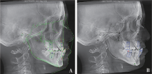
Fig.1
Soft and hard tissue anatomical landmark positioning and measurement"

Tab.1
Definition of anatomical landmarks related to soft and hard tissue measurements"
| 标记点 | 定义 |
|---|---|
| 蝶鞍点(S) | 蝶鞍影像的中心点 |
| 鼻根点(N) | 鼻额缝的最前点 |
| 耳点(P) | 外耳道的最上点 |
| 眶点(O) | 眶下缘的最低点 |
| 颧弓点(ZA) | 为颧弓三角前部之尖点 |
| 上牙槽座点(A) | 前鼻嵴与上牙槽缘点间的骨部最凹点 |
| 下牙槽座点(B) | 下牙槽缘点与颏前点间的骨部最凹点 |
| 前鼻棘点(ANS) | 前鼻棘之尖点 |
| 后鼻棘点(PNS) | 硬腭后部骨棘之尖点 |
| 颏前点(Po) | 颏部之最突点 |
| 颏下点(Me) | 颏部之最下点 |
| 颏顶点(Gn) | 颏前点与颏下点之中点 |
| 软组织鼻根点(Ns) | 软组织侧面上相应的鼻根点 |
| 颏唇沟点(Bs) | 颏唇沟最凹点 |
| 上唇突点(UL) | 上唇的最突点 |
| 下唇突点(LL) | 下唇的最突点 |
| 眶耳平面(FH) | 经过眶点和耳点的连接平面 |
| 腭平面(PP) | 经过前鼻棘点和后鼻棘点的连接平面 |
| 下颌平面(MP) | 通过颏下点与下颌角下缘相切的线 |
| 审美平面(EP) | 通过鼻尖和颏部最突点的切线 |
Tab.2
Soft and hard tissue measurement items"
| 测量项目 | 定义 |
|---|---|
| SNA/(°) | 由蝶鞍中心-鼻根点-上牙槽座点连线所构成角 |
| SNB/(°) | 由蝶鞍中心-鼻根点-下牙槽座点连线所构成角 |
| ANB/(°) | 由上牙槽座点-鼻根点-下牙槽座点连线所构成角 |
| MP-FH/(°) | 即下颌平面角,由下颌平面与眶耳平面的交角 |
| SN-PP/(°) | 前颅底平面与腭平面的交角 |
| SN-MP/(°) | 前颅底平面与下颌平面的交角 |
| Y轴角/(°) | 蝶鞍中心与颏顶点的连线与眶耳平面相交的下前角 |
| U1-NA/(°) | 上中切牙长轴与鼻根点-上牙槽座点连线交角 |
| U1-SN/(°) | 上中切牙长轴与前颅底平面相交的下内角 |
| L1-NB/(°) | 下中切牙长轴与鼻根点-下牙槽座点连线的交角 |
| L1-MP/(°) | 下中切牙长轴与下颌平面相交的上内角 |
| U1-L1/(°) | 上下中切牙长轴的交角 |
| S-Ns-Sn/(°) | 即上颌软组织突度,由蝶鞍点-软组织鼻根点-鼻下点连线交角 |
| S-Ns-Bs/(°) | 即下颌软组织突度,由蝶鞍点-软组织鼻根点-颏唇沟点连线交角 |
| Sn-Ns-Bs/(°) | 即上下颌软组织突度差的角度 |
| UL-EP/mm | 上唇突点与审美平面的距离 |
| LL-EP/mm | 下唇突点与审美平面的距离 |
| LL-UL/mm | 上下唇突度差的距离 |
| A点前移量/mm | 上牙槽座点前移量,即治疗前后A点的水平距离 |
| B点后移量/mm | 下牙槽座点后移量,即治疗前后B点的水平距离 |
| ANS点前移量/mm | 前鼻棘点前移量,即治疗前后ANS点的水平距离 |
| N-ANS/mm | 从鼻根点至前鼻棘点的距离,即上面高度 |
| ANS-Me/mm | 从前鼻棘点至颏下点的距离,即下面高度 |
| U1-NA/mm | 上中切牙长轴与鼻根点-上牙槽座点连线的垂直距离 |
| L1-NB/mm | 下中切牙长轴与鼻根点-下牙槽座点连线的垂直距离 |
| 前牙覆盖/mm | 上下中切牙切缘的水平距离 |
| Wits值/mm | 上下牙槽座点AB向功能性𬌗平面作垂线,两垂足分别为Ao点和Bo点,然后测量Ao点与Bo点间的距离为Wits值 |

Fig.2
3D reconstruction before and after treatment"
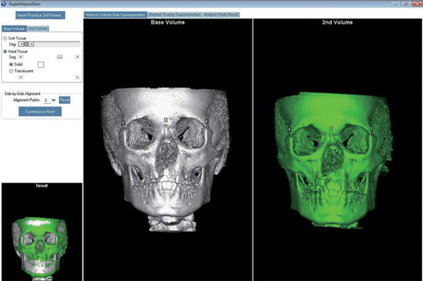
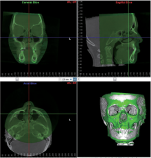
Fig.3
3D reconstruction and image overlap before and after treatment"

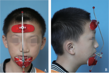
Fig.4
Maxillary anterior traction treatment"
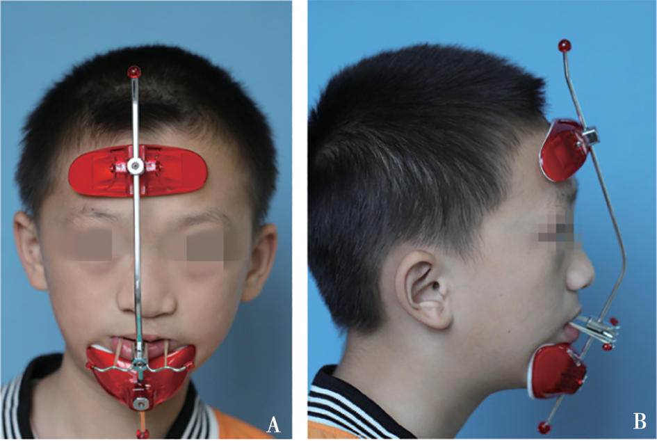
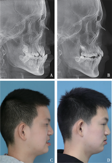
Fig.5
Lateral cephalometric radiographs and profiles before and after treatment"

Tab.3
Comparison of bone changes before and after treatment (sagittal direction)"
| 测量项目 | 治疗前(T1) | 治疗后(T2) | T2-T1 | P |
|---|---|---|---|---|
| SNA/(°) | 75.10±3.75 | 76.49±3.98 | 1.38±1.00 | 0.000* |
| SNB/(°) | 76.55±4.97 | 75.87±5.16 | -0.69±1.27 | 0.026* |
| ANB/(°) | -1.45±2.28 | 0.62±2.56 | 2.07±1.22 | 0.000* |
| Y轴角/(°) | 61.70±4.72 | 62.42±5.01 | 0.72±1.15 | 0.012* |
| ANS点前移量/mm | — | — | 1.65±0.61 | 0.000* |
| A点前移量/mm | — | — | 1.64±0.66 | 0.000* |
| B点后移量/mm | — | — | 1.63±1.34 | 0.000* |
Tab.4
Comparison of bone changes before and after treatment (vertical direction)"
| 测量项目 | 治疗前(T1) | 治疗后(T2) | T2-T1 | P |
|---|---|---|---|---|
| MP-FH/(°) | 29.68±5.35 | 31.14±5.96 | 1.47±1.74 | 0.001* |
| MP-SN/(°) | 39.72±6.24 | 41.09±6.21 | 1.38±2.30 | 0.015* |
| SN-PP/(°) | 16.32±6.28 | 14.11±6.65 | -2.22±2.74 | 0.002* |
| N-ANS/mm | 53.07±3.50 | 53.18±3.95 | 0.11±1.35 | 0.719 |
| ANS-Me/mm | 66.65±6.60 | 70.11±7.14 | 3.47±2.58 | 0.000* |
Tab.5
Comparison of dental changes before and after treatment"
| 测量项目 | 治疗前(T1) | 治疗后(T2) | T2-T1 | P |
|---|---|---|---|---|
| U1-NA/(°) | 19.59±10.02 | 22.93±7.93 | 3.34±4.69 | 0.005* |
| U1-NA/mm | 1.99±3.17 | 4.14±2.43 | 2.16±1.75 | 0.000* |
| U1-SN/(°) | 94.64±10.57 | 99.52±8.88 | 4.88±4.81 | 0.000* |
| L1-NB/(°) | 15.65±7.82 | 12.91±7.66 | -2.74±3.19 | 0.001* |
| L1-NB/mm | 3.80±2.63 | 2.43±2.64 | -1.38±1.39 | 0.000* |
| L1-MP/(°) | 79.56±8.57 | 76.18±8.56 | -3.38±3.45 | 0.000* |
| U1-L1/(°) | 145.87±13.92 | 144.94±12.13 | -0.94±6.64 | 0.537 |
| 前牙覆盖/mm | -3.75±3.50 | 4.08±2.67 | 7.83±2.14 | 0.000* |
| Wits值 | -2.29±4.05 | 2.98±3.87 | 5.27±1.96 | 0.000* |
Tab.6
Comparison of soft tissue changes before and after treatment"
| 测量项目 | 治疗前(T1) | 治疗后(T2) | T2-T1 | P |
|---|---|---|---|---|
| S-Ns-Sn/(°) | 86.01±4.37 | 88.29±3.61 | 2.29±1.73 | 0.000* |
| S-Ns-Bs/(°) | 85.68±5.40 | 85.80±4.93 | 0.13±2.08 | 0.791 |
| Sn-Ns-Bs/(°) | 0.63±3.51 | 2.49±3.21 | 1.86±2.88 | 0.009* |
| UL-EP/mm | -2.96±2.08 | -0.44±1.52 | 2.52±1.29 | 0.000* |
| LL-EP/mm | 2.40±2.20 | 2.16±1.82 | -0.24±1.45 | 0.477 |
| LL-UL/mm | -5.35±2.95 | -2.60±2.19 | 2.76±1.60 | 0.000* |
| [1] | Tan CS, Hariri F, Hassan MK. Severe midface and maxillary hypoplasia in non-cleft and non-syndromic patients: A 2-stage surgical strategy using distraction osteogenesis and orthognathic surgery[J]. J Stomatol Oral Maxillofac Surg, 2024, 125(3): 101552. |
| [2] | Cumerlato CBDF, Santos CSD, Santos MBFD, et al. Multidisciplinary approach for reestablishing function and aesthetic of unilateral cleft lip defect: A case report[J]. Cleft Palate Craniofac J, 2021, 58(3): 396-399. |
| [3] |
王斌卿, 宋涛. 青少年唇腭裂上颌后缩畸形治疗方法研究进展[J]. 中国实用口腔科杂志, 2020, 13(2): 117-122.
doi: 10.19538/j.kq.2020.02.012 |
| [4] |
Yamagata K, Mohri T, Watanabe A, et al. Anterior maxillary distraction osteogenesis with bone-borne intraoral buccal devices for maxillary hypoplasia with cleft lip and palate[J]. J Craniofac Surg, 2023, 34(6): 1867-1871.
doi: 10.1097/SCS.0000000000009412 pmid: 37253151 |
| [5] | Nys M, Bempt MVD, Shaheen E, et al. Three-dimensional planning accuracy and follow-up of Le Fort Ⅰ osteotomy in cleft lip/palate patients[J]. J Stomatol Oral Maxillofac Surg, 2023, 124(4): 101421. |
| [6] | 普盼君, 赵华翔, 牟清楠, 等. 唇腭裂上颌后缩的序列治疗[J]. 口腔医学, 2024, 44(8): 570-575. |
| [7] | 李巍然. 唇腭裂序列治疗中的正畸治疗[J]. 中华口腔医学杂志, 2023, 58(9):877-881. |
| [8] |
邹淑娟, 尹星, 周陈晨. 唇腭裂综合序列治疗中的牙颌畸形矫治[J]. 口腔疾病防治, 2020, 28(11): 681-688.
doi: 10.12016/j.issn.2096-1456.2020.11.001 |
| [9] | 赵飘, 陈泽策, 廖成成, 等. 前方牵引治疗的不同方式及比较[J]. 北京口腔医学, 2023, 31(3): 225-228. |
| [10] | 候任亚, 张鑫, 郭杰. 前方牵引治疗早期骨性Ⅲ类错𬌗的研究进展[J]. 中华口腔正畸学杂志, 2019, 26(1): 43-47. |
| [11] | Li RM, Shan YH, Li YF, et al. Zygomaticomaxillary suture maturation evaluation in patients with and without cleft lip and palate[J]. Am J Orthod Dentofacial Orthop, 2022, 162(2): 162-172. |
| [12] |
Angelieri F, Ruellas AC, Yatabe MS, et al. Zygomaticomaxillary suture maturation: Part Ⅱ-The influence of sutural maturation on the response to maxillary protraction[J]. Orthod Craniofac Res, 2017, 20(3): 152-163.
doi: 10.1111/ocr.12191 pmid: 28660731 |
| [13] | 周孙欣, 霍娜, 李帅臣, 等. 改良上颌前方牵引对生长发育高峰后期上颌发育不足患者软组织侧貌的影响[J]. 实用口腔医学杂志, 2024, 40(3): 365-370. |
| [14] | 任冬宜. 快速扩弓合并前方牵引矫治上颌发育不足的临床研究[J]. 中国中西医结合耳鼻咽喉科杂志, 2022, 30(4): 280-282. |
| [15] |
Buschang PH, Porter C, Genecov E, et al. Face mask therapy of preadolescents with unilateral cleft lip and palate[J]. Angle Orthod, 1994, 64(2): 145-150.
pmid: 8010523 |
| [16] | So LL. Effects of reverse headgear treatment on sagittal correction in girls born with unilateral complete cleft lip and cleft palate: Skeletal and dental changes[J]. Am J Orthod Dentofacial Orthop, 1996, 109(2): 140-147. |
| [17] | Ross RB. Treatment variables affecting facial growth in complete unilateral cleft lip and palate[J]. Cleft Palate J, 1987, 24(1): 5-77. |
| [18] | Zhang Y, Jia H, Fu Z, et al. Dentoskeletal effects of facemask therapy in skeletal Class Ⅲ cleft patients with or without bone graft[J]. Am J Orthod Dentofacial Orthop, 2018, 153(4): 542-549. |
| [19] |
Dogan S. The effects of face mask therapy in cleft lip and palate patients[J]. Ann Maxillofac Surg, 2012, 2(2): 116-120.
doi: 10.4103/2231-0746.101332 pmid: 23483763 |
| [20] | Baccetti T, Franchi L, McNamara JA. Treatment and posttreatment craniofacial changes after rapid maxillary expansion and facemask therapy[J]. Am J Orthod Dentofac Orthop, 2000, 118(4): 404-413. |
| [21] |
Zhang D, Zheng L, Wang Q, et al. Displacements prediction from 3D finite element model of maxillary protraction with and without rapid maxillary expansion in a patient with unilateral cleft palate and alveolus[J]. Biomed Eng Online, 2015, 14: 80.
doi: 10.1186/s12938-015-0074-9 pmid: 26285822 |
| [22] |
Ngan PW, Hagg U, Yiu C, et al. Treatment response and long-term dentofacial adaptations to maxillary expansion and protraction[J]. Semin Orthod, 1997, 3(4): 255-264.
doi: 10.1016/s1073-8746(97)80058-8 pmid: 9573887 |
| [23] | da Luz Vieira G, de Menezes LM, de Lima EMS, et al. Dentoskeletal effects of maxillary protraction in cleft patients with repetitive weekly protocol of alternate rapid maxillary expansions and constrictions[J]. Cleft Palate Craniofac J, 2009, 46(4): 391-398. |
| [24] | Kim JH, Viana MA, Graber TM, et al. The effectiveness of protraction face mask therapy: A meta-analysis[J]. Am J Orthod Dentofacial Orthop, 1999, 115(6): 675-685. |
| [25] | Foersch M, Jacobs C, Wriedt S, et al. Effectiveness of maxillary protraction using facemask with or without maxillary expansion: A systematic review and meta-analysis[J]. Clin Oral Investig, 2015, 19(6): 1181-1192. |
| Viewed | ||||||
|
Full text |
|
|||||
|
Abstract |
|
|||||