
口腔医学 ›› 2026, Vol. 46 ›› Issue (3): 181-188.doi: 10.13591/j.cnki.kqyx.2026.03.004
王潇辰1,2,3, 林君彦1,2,3, 郭培培1,2,3, 张蕊1,2,3, 李丹丹1,2,3( )
)
收稿日期:2025-11-01
出版日期:2026-03-28
发布日期:2026-03-31
基金资助:
WANG Xiaochen1,2,3, LIN Junyan1,2,3, GUO Peipei1,2,3, ZHANG Rui1,2,3, LI Dandan1,2,3()
Received:2025-11-01
Online:2026-03-28
Published:2026-03-31
摘要:
目的 探究低水平激光治疗(low-level laser therapy,LLLT)对正畸牙齿移动速度的影响及机制。方法 构建大鼠牙移动与低水平激光治疗模型,通过Micro-CT比较LLLT辅助正畸加力组(F+L+组)大鼠与单纯正畸加力组(F+L-组)大鼠第一磨牙近中移动距离。构建THP-1细胞压力与激光照射模型,通过mRNA测序分析F+L+组和F+L-组THP-1细胞的差异表达基因并进行富集分析,并通过qRT-PCR、蛋白免疫印迹实验测定通路相关基因及标志物的表达变化。结果 F+L+组大鼠上颌第一磨牙近中移动距离比F+L-组显著增加(P<0.05);F+L+组THP-1相较F+L-组THP-1差异基因显著富集于线粒体自噬通路,qRT-PCR及蛋白免疫印迹实验验证F+L+组THP-1的线粒体自噬相关基因OPTN、SQSTM1表达水平较F+L-组THP-1降低,同时其线粒体自噬通路关键标志物PINK1/Parkin也下降。结论 低水平激光治疗可能通过抑制线粒体自噬通路加速正畸牙移动。
中图分类号:
王潇辰, 林君彦, 郭培培, 张蕊, 李丹丹. 低水平激光通过线粒体自噬通路促进正畸牙移动的作用研究[J]. 口腔医学, 2026, 46(3): 181-188.
WANG Xiaochen, LIN Junyan, GUO Peipei, ZHANG Rui, LI Dandan. Effect of low-level laser therapy on orthodontic tooth movement via the mitophagy pathway[J]. Stomatology, 2026, 46(3): 181-188.
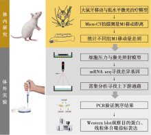
图1
低水平激光通过线粒体自噬通路促进大鼠正畸牙移动的效应研究技术路线图"
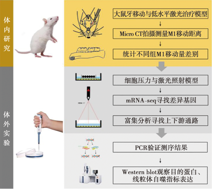

图2
大鼠牙移动与低水平激光治疗模型构建 A:大鼠LLLT辅助牙移动模型构建示意图;B:大鼠口内操作图;C:LLLT辅助牙移动实验策略图。"
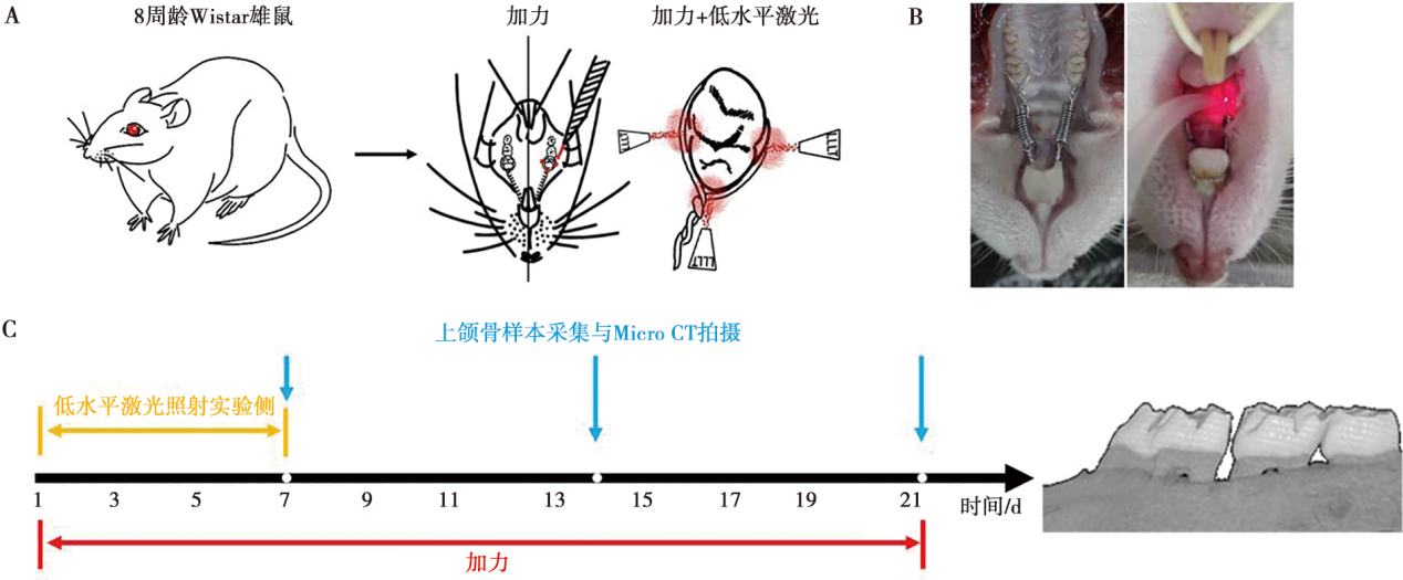
表1
qRT-PCR所用引物序列"
| 基因名称 | 引物序列 |
|---|---|
| SQSTM1 | F:5'-GACTACGACTTGTGTAGCGTC-3',R:5'-AGTGTCCGTGTTTCACCTTCC-3' |
| SRC | F:5'-GAGCGGCTCCAGATTGTCAA-3',R:5'-CTGGGGATGTAGCCTGTCTGT-3' |
| TFEB | F:5'-ACCTGTCCGAGACCTATGGG-3',R:5'-CGTCCAGACGCATAATGTTGTC-3' |
| GABARAPL1 | F:5'-ATGAAGTTCCAGTACAAGGAGGA-3',R:5'-GCTTTTGGAGCCTTCTCTACAAT-3' |
| HIF1A | F:5'-GAACGTCGAAAAGAAAAGTCTCG-3',R:5'-CCTTATCAAGATGCGAACTCACA-3' |
| OPTN | F:5'-AAAGAGCGTCTAATGGCCTTG-3',R:5'-GTTCAGACACGATGCCCAACA-3' |
| RELA | F:5'-ATGTGGAGATCATTGAGCAGC-3',R:5'-CCTGGTCCTGTGTAGCCATT-3' |
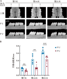
图3
低水平激光处理后大鼠上颌骨第一磨牙移动距离 A:第7、14、21天F+L-大鼠与F+L+大鼠上颌骨样本Micro-CT第一磨牙牙齿移动情况;B:牙齿移动距离统计图;OTM:牙移动方向;**:P<0.01;***:P<0.001。"
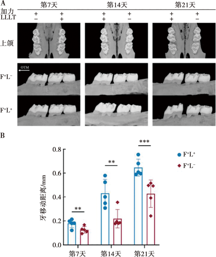
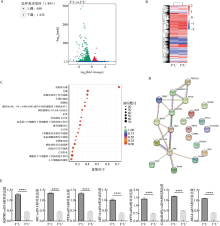
图4
低水平激光加力后THP-1细胞mRNA测序结果 A:F+L+和F+L-THP-1细胞mRNA测序差异表达基因火山图;B:F+L+和F+L-THP-1细胞差异表达基因聚类分析热图;C:F+L+和F+L-THP-1细胞差异表达基因KEGG富集分析气泡图;D:富集在线粒体自噬通路的基因PPI网络分析图;E:线粒体自噬通路PPI提示存在互作的基因在F+L-和F+L+ THP-1细胞差异表达基因mRNA相对表达量;F+L-:加力组;F+L+:加力辅助激光照射组;****:P<0.000 1。"

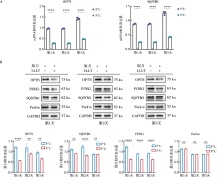
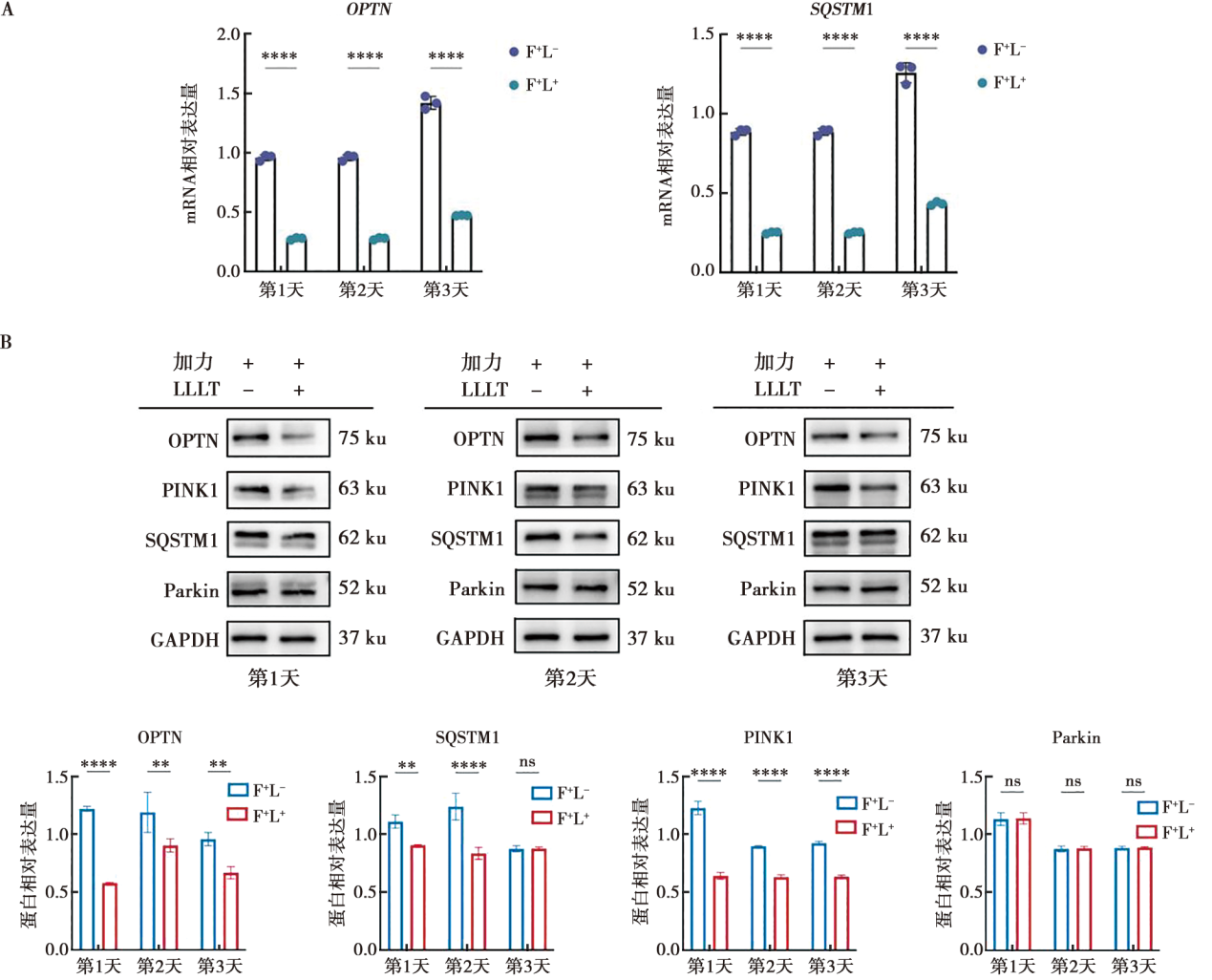
图5
低水平激光下调THP-1细胞线粒体自噬通路 A:在THP-1细胞处理1、2、3 d后,对线粒体自噬通路上的两个关键受体OPTN、SQSTM1的表达量进行PCR检测;B:在THP-1细胞处理1、2、3 d后,对线粒体自噬通路关键受体、marker基因表达量进行Western blot检测;F+L-:加力组;F+L+:加力辅助激光照射组;**:P<0.01;****:P<0.000 1;ns:P>0.05。"
| [1] |
Ren YJ, Maltha JC, Kuijpers-Jagtman AM. Optimum force magnitude for orthodontic tooth movement: A systematic literature review[J]. Angle Orthod, 2003, 73(1): 86-92.
doi: 10.1043/0003-3219(2003)073<0086:OFMFOT>2.0.CO;2 pmid: 12607860 |
| [2] |
Yassir YA, McIntyre GT, Bearn DR. Orthodontic treatment and root resorption: An overview of systematic reviews[J]. Eur J Orthod, 2021, 43(4): 442-456.
doi: 10.1093/ejo/cjaa058 |
| [3] |
Villaman-Santacruz H, Torres-Rosas R, Acevedo-Mascarúa AE, et al. Root resorption factors associated with orthodontic treatment with fixed appliances: A systematic review and meta-analysis[J]. Dent Med Probl, 2022, 59(3): 437-450.
doi: 10.17219/dmp/145369 |
| [4] |
Baghizadeh Fini M, Olyaee P, Homayouni A. The effect of low-level laser therapy on the acceleration of orthodontic tooth movement[J]. J Lasers Med Sci, 2020, 11(2): 204-211.
doi: 10.34172/jlms.2020.34 pmid: 32273964 |
| [5] |
Hamblin MR. Mechanisms and applications of the anti-inflammatory effects of photobiomodulation[J]. AIMS Biophys, 2017, 4(3): 337-361.
doi: 10.3934/biophy.2017.3.337 |
| [6] |
Escobar LM, Grajales M, Bendahan Z, et al. Laser-stimulated human gingival fibroblasts: Alterations in migration, secretome production, and induction of reactive oxygen species[J]. Lasers Med Sci, 2025, 40(1): 266.
doi: 10.1007/s10103-025-04499-4 |
| [7] |
Luo ZQ, He YQ, Wu HK, et al. Efficacy of laser adjuvant therapy in the management of post-operative endodontic pain: A systematic review and meta-analysis[J]. Int Endod J, 2024, 57(12): 1700-1716.
doi: 10.1111/iej.14140 pmid: 39287434 |
| [8] |
Zou QR, Zhang SX, Jiang CW, et al. Low-level laser therapy on soft tissue healing after implantation: A randomized controlled trial[J]. BMC Oral Health, 2024, 24(1): 1477.
doi: 10.1186/s12903-024-05258-7 pmid: 39639276 |
| [9] |
Carneiro AMP, Barros APO, de Oliveira RP, et al. The effect of photobiomodulation using low-level laser therapy on tooth sensitivity after dental bleaching: A systematic review[J]. Lasers Med Sci, 2022, 37(7): 2791-2804.
doi: 10.1007/s10103-022-03578-0 |
| [10] |
Ali Al-Maweri S, Javed F, Kalakonda B, et al. Efficacy of low level laser therapy in the treatment of burning mouth syndrome: A systematic review[J]. Photodiagnosis Photodyn Ther, 2017, 17: 188-193.
doi: S1572-1000(16)30253-8 pmid: 27919663 |
| [11] |
Grajales M, Ríos-Osorio N, Jimenez-Peña O, et al. Effectiveness of photobiomodulation with low-level lasers on the acceleration of orthodontic tooth movement: A systematic review and meta-analysis of split-mouth randomised clinical trials[J]. Lasers Med Sci, 2023, 38(1): 200.
doi: 10.1007/s10103-023-03870-7 |
| [12] | Caccianiga G, Giudice AL, Longoni S, et al. Low-level laser therapy protocols in dental movement acceleration and in pain management during orthodontic treatment[J]. J Biol Regul Homeost Agents, 2019, 33(6 Suppl. 1): 59-68. |
| [13] |
Yong JW, Gröger S, VON Bremen J, et al. Photobiomodulation therapy assisted orthodontic tooth movement: Potential implications, challenges, and new perspectives[J]. J Zhejiang Univ Sci B, 2023, 24(11): 957-973.
doi: 10.1631/jzus.B2200706 |
| [14] | 戴佳韵, 王姝婧, 林君彦, 等. 低水平激光用于加速正畸牙齿移动的研究进展[J]. 口腔医学, 2022, 42(12): 1123-1128. |
| [15] | 时之凯, 王斌, 韩爽, 等. 低能量激光治疗加速正畸牙齿移动及减轻疼痛的临床研究[J]. 实用口腔医学杂志, 2025, 41(2): 235-238. |
| [16] | El-Angbawi A, McIntyre G, Fleming PS, et al. Non-surgical adjunctive interventions for accelerating tooth movement in patients undergoing orthodontic treatment[J]. Cochrane Database Syst Rev, 2023, 6(6): CD010887. |
| [17] |
Dominguez A. Current protocol to achieve dental movement acceleration and pain control with Photo-biomodulation[J]. World J Methodol, 2023, 13(5): 379-383.
doi: 10.5662/wjm.v13.i5.379 |
| [18] | Ali Karabel M, Doğru M, Doğru A, et al. Evaluation of the effects of diode laser application on experimental orthodontic tooth movements in rats. Histopathological analysis[J]. Acta Cir Bras, 2021, 35(12): e351204. |
| [19] |
Nakai Y, Praneetpong N, Ono W, et al. Mechanisms of osteoclastogenesis in orthodontic tooth movement and orthodontically induced tooth root resorption[J]. J Bone Metab, 2023, 30(4): 297-310.
doi: 10.11005/jbm.2023.30.4.297 |
| [20] |
Alghamdi B, Jeon HH, Ni J, et al. Osteoimmunology in periodontitis and orthodontic tooth movement[J]. Curr Osteoporos Rep, 2023, 21(2): 128-146.
doi: 10.1007/s11914-023-00774-x pmid: 36862360 |
| [21] | Wang HN, Shen YQ. MicroRNA-20a negatively regulates the growth and osteoclastogenesis of THP-1 cells by downregulating PPARγ[J]. Mol Med Rep, 2019, 20(5): 4271-4276. |
| [22] |
Li S, Li Q, Zhu Y, et al. GDF15 induced by compressive force contributes to osteoclast differentiation in human periodontal ligament cells[J]. Exp Cell Res, 2020, 387(1): 111745.
doi: 10.1016/j.yexcr.2019.111745 |
| [23] |
Deng J, Zhuang ZM, Xu X, et al. Mechanical force increases tooth movement and promotes remodeling of alveolar bone defects augmented with bovine bone mineral[J]. Prog Orthod, 2024, 25(1): 2.
doi: 10.1186/s40510-023-00501-3 pmid: 38185724 |
| [24] |
Tan K, Wang JY, Su XY, et al. KAT6A/YAP/TEAD4 pathway modulates osteoclastogenesis by regulating the RANKL/OPG ratio on the compression side during orthodontic tooth movement[J]. Prog Orthod, 2024, 25(1): 29.
doi: 10.1186/s40510-024-00530-6 pmid: 39129034 |
| [25] |
Funaki-Dohi M, Hotokezaka Y, Hotokezaka H, et al. Importance of the early phase of orthodontic force application in the induction of root resorption[J]. Angle Orthod, 2025, 95(3): 323-331.
doi: 10.2319/060324-433.1 pmid: 39909061 |
| [26] |
Nogueira AVB, Marcantonio CC, de Molon RS, et al. Experimental models of orthodontic tooth movement and their effects on periodontal tissues remodelling[J]. Arch Oral Biol, 2021, 130: 105216.
doi: 10.1016/j.archoralbio.2021.105216 |
| [27] |
Cadenas de Llano-Pérula M, Zong C, Van Dessel J, et al. 3D quantification of orthodontic tooth movement in rats by means of micro-computed tomography[J]. Clin Oral Investig, 2022, 26(5): 3911-3920.
doi: 10.1007/s00784-021-04358-w |
| [28] |
Baser Keklikci H, Yagci A. Effects of different wavelengths of low-level laser therapy on orthodontically induced inflammatory root resorption in rats investigated with micro-computerized tomography[J]. Am J Orthod Dentofac Orthop, 2021, 159(3): e245-e251.
doi: 10.1016/j.ajodo.2020.10.020 |
| [29] |
Baser Keklikci H, Yagci A, Yay AH, et al. Effects of 405-, 532-, 650-, and 940-nm wavelengths of low-level laser therapies on orthodontic tooth movement in rats[J]. Prog Orthod, 2020, 21(1): 43.
doi: 10.1186/s40510-020-00343-3 pmid: 33258041 |
| [30] |
Vanderlei BMC, Torres MC, Paredes N, et al. Effect of photobiomodulation and corticopuncture methods on tooth displacement and gene expression: Animal study[J]. Lasers Med Sci, 2024, 39(1): 283.
doi: 10.1007/s10103-024-04136-6 |
| [31] |
An Y, Zhao JM, Xu H, et al. Effect of humic acid as a photosensitizer combined with low-energy laser on orthodontic tooth movement in rats[J]. J Dent Sci, 2022, 17(1): 407-414.
doi: 10.1016/j.jds.2021.08.006 pmid: 35028064 |
| [32] |
Nakatani A, Kunimatsu R, Tsuka Y, et al. Effects of high-frequency near infrared laser irradiation on experimental tooth movement-induced pain in rats[J]. Lasers Med Sci, 2022, 37(6): 2697-2706.
doi: 10.1007/s10103-022-03543-x |
| [33] |
D’Arcy MS. Mitophagy in health and disease. Molecular mechanisms, regulatory pathways, and therapeutic implications[J]. Apoptosis, 2024, 29(9/10): 1415-1428.
doi: 10.1007/s10495-024-01977-y |
| [34] |
Abulaiti G, Qin X, Chen LL, et al. Mitophagy and its significance in periodontal disease[J]. Oral Dis, 2025, 31(7): 2001-2018.
doi: 10.1111/odi.v31.7 |
| [35] |
Gou H, Wang T, Chen Y, et al. Role of Pink1 in regulating osteoclast differentiation during periodontitis[J]. J Dent Res, 2025, 104(7): 753-762.
doi: 10.1177/00220345251315723 pmid: 40075549 |
| [36] | Jang JS, Hong SJ, Mo SZ, et al. PINK1 restrains periodontitis-induced bone loss by preventing osteoclast mitophagy impairment[J]. Redox Biol, 2024, 69: 103023. |
| [37] |
Liu BQ, Zhang J, Liu GJ, et al. Expression of PINK1 and Parkin in human apical periodontitis[J]. Int Endod J, 2022, 55(8): 870-881.
doi: 10.1111/iej.13760 pmid: 35502680 |
| [38] |
Bharath LP, Agrawal M, McCambridge G, et al. Metformin enhances autophagy and normalizes mitochondrial function to alleviate aging-associated inflammation[J]. Cell Metab, 2020, 32(1): 44-55. e6.
doi: S1550-4131(20)30197-2 pmid: 32402267 |
| [39] |
Liu RX, Xu CL, Zhang WL, et al. FUNDC1-mediated mitophagy and HIF1α activation drives pulmonary hypertension during hypoxia[J]. Cell Death Dis, 2022, 13(7): 634.
doi: 10.1038/s41419-022-05091-2 pmid: 35864106 |
| [40] | 陈佳锋, 傅修涛, 丁振斌. 自噬调控多功能蛋白p62/SQSTM1参与肿瘤及其微环境的研究进展[J]. 中国临床医学, 2020, 27(2): 321-326. |
| [41] | Lin CC, Yan J, Kapur MD, et al. Parkin coordinates mitochondrial lipid remodeling to execute mitophagy[J]. EMBO Rep, 2022, 23(12): e55191. |
| [42] | 王香香, 凌江红, 王煜姣, 等. Pink1/Parkin信号通路调控线粒体自噬的研究进展[J]. 基因组学与应用生物学, 2022, 41 (4): 919-926. |
| [43] |
Ryan TA, Tumbarello DA. Optineurin: A coordinator of membrane-associated cargo trafficking and autophagy[J]. Front Immunol, 2018, 9: 1024.
doi: 10.3389/fimmu.2018.01024 pmid: 29867991 |
| [44] | 范智博, 金珂, 李胜鸿, 等. 饥饿条件下活性氧通过PINK1/Parkin通路调控人牙周膜细胞的线粒体自噬[J]. 华西口腔医学杂志, 2022, 40(6): 645-653. |
| [45] |
Clark IE, Dodson MW, Jiang CG, et al. Drosophila pink1 is required for mitochondrial function and interacts genetically with parkin[J]. Nature, 2006, 441(7097): 1162-1166.
doi: 10.1038/nature04779 |
| [46] | 张明军, 罗俊一, 杨毅宁. PINK1、Parkin与泛素协同调控线粒体自噬的研究进展[J]. 医学综述, 2020, 26(10): 1887-1892. |
| [47] |
Qiu YP, Wang JC, Li H, et al. Emerging views of OPTN(optineurin)function in the autophagic process associated with disease[J]. Autophagy, 2022, 18(1): 73-85.
doi: 10.1080/15548627.2021.1908722 |
| [48] |
Lazarou M, Sliter DA, Kane LA, et al. The ubiquitin kinase PINK1 recruits autophagy receptors to induce mitophagy[J]. Nature, 2015, 524(7565): 309-314.
doi: 10.1038/nature14893 |
| [49] |
Wang RM, Dong Y, Lu YJ, et al. Photobiomodulation for global cerebral ischemia: Targeting mitochondrial dynamics and functions[J]. Mol Neurobiol, 2019, 56(3): 1852-1869.
doi: 10.1007/s12035-018-1191-9 pmid: 29951942 |
| [50] |
Lu YJ, Wang RM, Dong Y, et al. Low-level laser therapy for beta amyloid toxicity in rat hippocampus[J]. Neurobiol Aging, 2017, 49: 165-182.
doi: S0197-4580(16)30243-3 pmid: 27815990 |
| [1] | 王若雯, 王华. 下颌切牙牙根与牙槽骨反应的差异——骨性Ⅲ类掩饰性与联合治疗比较[J]. 口腔医学, 2026, 46(5): 363-368. |
| [2] | 钟悦, 吕丹, 张译尹, 黄子铭, 张元, 谢理哲, 钱雅婧. 基于3dMD的正畸治疗后成人面部软组织变化的研究[J]. 口腔医学, 2026, 46(4): 283-288. |
| [3] | 段沛沛, 余芯乐, 王韵诗, 韩向龙. 微信公众号赋能《口腔正畸学》本科生实践教学的探索与思考[J]. 口腔医学, 2025, 45(8): 619-623. |
| [4] | 陈子昂, 刘子昂, 欧阳雨晴, 娄依婷, 施洁珺, 丁王辉. 正畸治疗中周围解剖结构与牙根外吸收之间关系的综述[J]. 口腔医学, 2025, 45(5): 394-400. |
| [5] | 方铁钧, 梁惠敏, 黄敏莉. 带牵引设计的铸造桩核冠向牵引龈下牙折上前牙的临床效果[J]. 口腔医学, 2025, 45(4): 275-280. |
| [6] | 韩陆琦, 沈慧婕, 陆山鸣, 李国情, 王威, 汤欢. 下颌切牙先天缺失患者间隙关闭前后的骨量变化[J]. 口腔医学, 2025, 45(12): 909-913. |
| [7] | 王宇婷, 胡敏, 范炜, 顾启慧, 王震东, 朱琳琳. 下颌第三磨牙生长及萌出情况与下颌牙列拥挤度的相关性研究[J]. 口腔医学, 2025, 45(11): 808-813. |
| [8] | 刘浩, 彭文静, 高秋颖, 胥加斌, 刘刚, 朱绍跃. PAOO微创术式与传统术式促进正畸牙移动效果及生物学机制研究[J]. 口腔医学, 2025, 45(11): 813-818. |
| [9] | 彭焱, 张驰, 高雳, 李希庭, 赵川江. Ⅳ期牙周炎患者的正畸考量和时机选择[J]. 口腔医学, 2025, 45(1): 25-36. |
| [10] | 包佳琦, 王中秀, 冯贻苗, 雷利红, 陈莉丽. 正畸治疗中牙周硬组织相关并发症的处理[J]. 口腔医学, 2025, 45(1): 37-44. |
| [11] | 周悦, 唐振兴, 李宇. 拔除前磨牙正畸病例的磨牙支抗丧失及其影响因素[J]. 口腔医学, 2025, 45(1): 64-68. |
| [12] | 张琳琳, 刘东旭. 骨性Ⅲ类偏颌畸形牙骨特征、分类及治疗相关研究进展[J]. 口腔医学, 2024, 44(9): 710-714. |
| [13] | 普盼君, 赵华翔, 牟清楠, 梁蓉, 侯玉霞. 唇腭裂上颌后缩的序列治疗[J]. 口腔医学, 2024, 44(8): 570-575. |
| [14] | 夏雪妍, 吴梦婕. 唇部形态影响因素及其与正畸治疗相关性的研究进展[J]. 口腔医学, 2024, 44(8): 635-640. |
| [15] | 宋野, 任银婷, 张轶涵, 韩晶莹. 不同拔牙方式对骨性Ⅱ类错牙合畸形Bolton指数及咬合关系的影响[J]. 口腔医学, 2024, 44(5): 334-337. |
| 阅读次数 | ||||||
|
全文 |
|
|||||
|
摘要 |
|
|||||
 苏公网安备32010602011670号