
Stomatology ›› 2023, Vol. 43 ›› Issue (10): 903-909.doi: 10.13591/j.cnki.kqyx.2023.10.008
• Clinical Research • Previous Articles Next Articles
CHENG Aoran1,WANG Jue2,FAN Yawei1,3( )
)
Revised:2023-04-16
Online:2023-10-28
Published:2023-10-20
CLC Number:
CHENG Aoran, WANG Jue, FAN Yawei. Analysis of morphological and morphometric characteristics of canalis sinuosus and its accessory canals: A cone-beam CT study[J]. Stomatology, 2023, 43(10): 903-909.
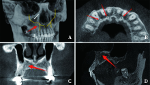
Fig.1
Schematic representation of the canalis sinuosus and its accessory canals"

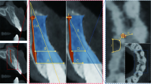
Fig.2
CBCT slices and measurement landmarks"

Tab.1
Detection of ACs in gender and age groups"
| 组别 | 检出 | 未检出 | P | |
|---|---|---|---|---|
| 性别 | ||||
| 男性 | 74 | 84 | 0.001 | 0.970 |
| 女性 | 83 | 95 | ||
| 年龄组 | ||||
| 18~37岁 | 77 | 91 | 2.267 | 0.322 |
| 38~57岁 | 65 | 63 | ||
| 58岁以上 | 15 | 25 |
Tab.2
Distribution of ACs in gender and age groups"
| 组别 | 仅左侧检出 (n=30) | 仅右侧检出 (n=53) | 双侧检出 (n=74) |
|---|---|---|---|
| 性别 | |||
| 男性 | 16 | 22 | 36 |
| 女性 | 14 | 31 | 38 |
| 年龄组 | |||
| 18~37岁 | 14 | 30 | 33 |
| 38~57岁 | 13 | 17 | 35 |
| 58岁以上 | 3 | 6 | 6 |

Fig.3
Accessory canals localization"
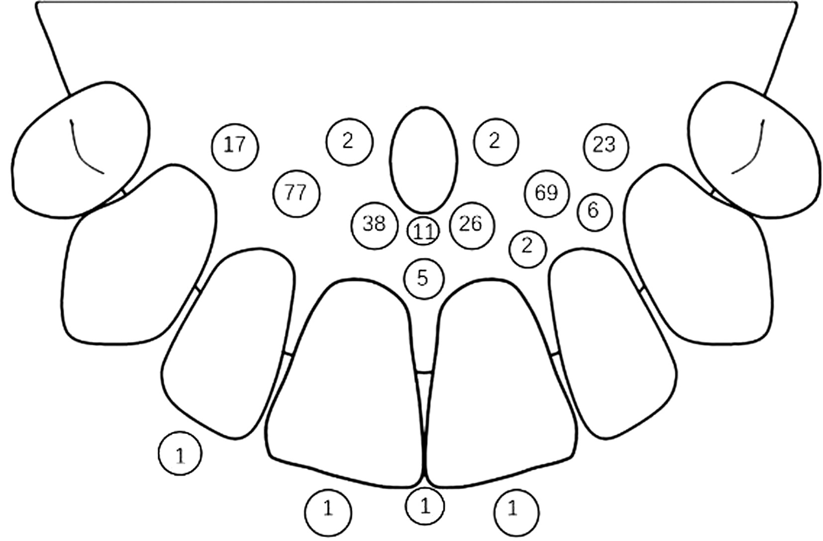
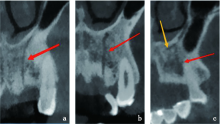
Fig.4
Schematic diagram of the ACs classification"

Tab.3
Summary of morphological characteristic parameters of CS and its ACs classified by gender and age groups"
| 组别 | A/mm | B/mm | C/mm | D/mm | E/mm | F/mm | ∠α/(°) | G/mm | H/mm | I/mm |
|---|---|---|---|---|---|---|---|---|---|---|
| 18~37岁 | 8.70(7.13,9.82) | 5.35±1.72 | 1.95(1.50,2.40) | 3.15(1.95,4.46) | 1.17±0.37 | 8.03±1.97 | 16.20(9.58,21.81) | 0.92(0.80,1.43) | 1.38±0.57 | 1.40(1.08,2.15) |
| 38~57岁 | 8.44(7.83,9.60) | 6.01±1.46 | 2.26(1.50,3.00) | 3.88(3.08,4.42) | 1.27±0.37 | 11.76±3.14a | 17.30(14.55,24.78) | 0.90(0.72,1.09) | 1.51±0.59 | 1.09(0.90,2.48) |
| 58岁以上 | 9.30(7.20,9.45) | 6.84±1.46 | 3.00(1.95,3.45) | 4.20(4.05,4.50) | 1.10±0.21 | 12.13±2.55a | 12.70(1.10,28.15) | 1.10(1.10,1.25) | 2.18±0.85ab | 0.80(0.50,2.40) |
| H/F | 0.080 | 2.102 | 2.886 | 4.167 | 0.722 | 13.063 | 1.047 | 2.421 | 3.456 | 1.461 |
| P | 0.961 | 0.134 | 0.236 | 0.125 | 0.491 | 0.000** | 0.592 | 0.298 | 0.041* | 0.482 |
| 男性 | 8.70(8.04,9.90) | 6.02±1.40 | 2.10(1.50,2.70) | 3.67±1.24 | 1.24±0.34 | 10.49±3.58 | 17.78±9.07 | 0.91(0.90,1.20) | 1.51±0.61 | 1.30(0.90,2.50) |
| 女性 | 8.04(6.60,9.38) | 5.48±1.93 | 2.40(1.95,3.23) | 3.65±0.79 | 1.16±0.38 | 9.64±2.58 | 17.56±5.75 | 0.92(0.70,1.20) | 1.57±0.71 | 1.15(0.85,2.18) |
| Z/t | -1.299 | 1.128 | -1.578 | 0.054 | 0.877 | 0.919 | 0.100 | -0.696 | -0.330 | -0.190 |
| P | 0.194 | 0.265 | 0.115 | 0.957 | 0.385 | 0.363 | 0.921 | 0.487 | 0.743 | 0.849 |
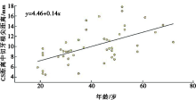
Fig.5
Correlation between age and the distance from CS to the root apex of the central incisor"

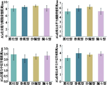
Fig.6
Comparison of morphological characteristics of ACs in patients with different NPC types"

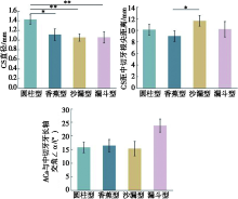
Fig.7
Comparison of morphological characteristic parameters of CS and ACs in patients with different NPC types"


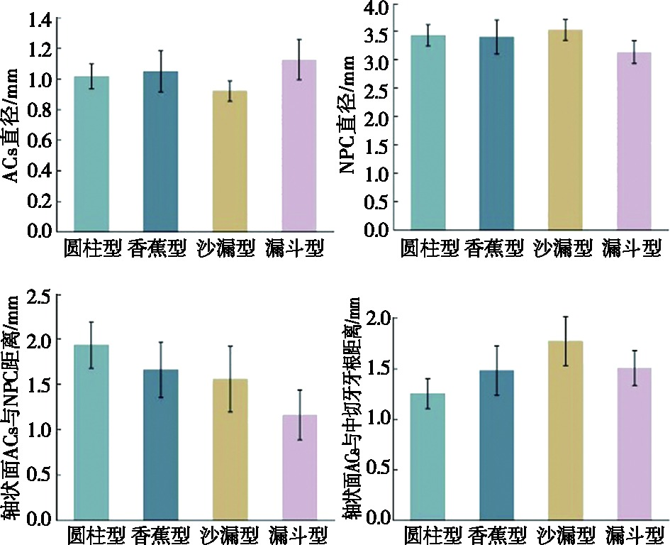
Fig.8
Comparison of ACs and NPC morphological characteristic parameters in patients with different NPC types"
| [1] | 李志进, 郭家平. 鼻腭管区种植的研究进展[J]. 中国口腔种植学杂志, 2019, 24(2):92-97. |
| [2] |
Botermans A, Lidén A, de Carvalho Machado V, et al. Immediate implant placement in the maxillary aesthetic zone:A cone beam computed tomography study[J]. J Clin Med, 2021, 10(24):5853.
doi: 10.3390/jcm10245853 |
| [3] | 俞琼, 黄廷贲, 杨国利. 原位取骨技术在上前牙种植中的应用[J]. 口腔医学, 2021, 41(7):649-653. |
| [4] |
Jones FW. The anterior superior alveolar nerve and vessels[J]. J Anat, 1939, 73(Pt 4):583-591.
pmid: 17104781 |
| [5] | Rusu MC, Iacov-Crǎiţoiu MM, Sǎndulescu M, et al. Constant features of the adult maxillary bone in the site of the premaxillary suture:The suturanotha, Macalister’s foramina, Parinaud’scanal, and the second angle of the canalis sinuosus of Wood Jones[J]. Rom J Morphol Embryol, 2019, 60(4):1097-1103. |
| [6] |
de Oliveira-Neto OB, Barbosa FT, de Lima FJC, et al. Prevalence of canalis sinuosus and accessory canals of canalis sinuosus on cone beam computed tomography:A systematic review and meta-analysis[J]. Int J Oral Maxillofac Surg, 2023, 52(1):118-131.
doi: 10.1016/j.ijom.2022.06.011 |
| [7] |
von Arx T, Lozanoff S, Sendi P, et al. Assessment of bone channels other than the nasopalatine canal in the anterior maxilla using limited cone beam computed tomography[J]. Surg Radiol Anat, 2013, 35(9):783-790.
doi: 10.1007/s00276-013-1110-8 pmid: 23539212 |
| [8] | Vasiljevic M, Milanovic P, Jovicic N, et al. Morphological and morphometric characteristics of anterior maxilla accessory canals and relationship with nasopalatine canal type-A CBCT study[J]. Diagnostics (Basel), 2021, 11(8):1510. |
| [9] |
Bliggenstorfer S, Chappuis V, von Arx T. Misinterpretation of a periapical radiograph:The canalis sinuosus mimicking a root resorption[J]. Swiss Dent J, 2021, 131(12):999-1005.
pmid: 34854290 |
| [10] | Milanovic P, Selakovic D, Vasiljevic M, et al. Morphological characteristics of the nasopalatine canal and the relationship with the anterior maxillary bone-a cone beam computed tomography study[J]. Diagnostics (Basel), 2021, 11(5):915. |
| [11] | Tomrukçu DN, Köse TE. Assesment of accessory branches of canalis sinuosus on CBCT images[J]. Med Oral Patol Oral Cir Bucal, 2020, 25(1):e124-e130. |
| [12] | Arruda JA, Silva P, Silva L, et al. Dental implant in the canalis sinuosus:A case report and review of the literature[J]. Case Rep Dent, 2017, 2017:4810123. |
| [13] | Ghandourah AO, Rashad A, Heiland M, et al. Cone-beam tomographic analysis of canalis sinuosus accessory intraosseous canals in the maxilla[J]. Ger Med Sci, 2017, 15:Doc20. |
| [14] |
Orhan K, Gorurgoz C, Akyol M, et al. An anatomical variant:Evaluation of accessory canals of the canalis sinuosus using cone beam computed tomography[J]. Folia Morphol (Warsz), 2018, 77(3):551-557.
doi: 10.5603/FM.a2018.0003 pmid: 29345719 |
| [15] |
de C Machado V, Chrcanovic BR, Felippe MB, et al. Assessment of accessory canals of the canalis sinuosus:A study of 1 000 cone beam computed tomography examinations[J]. Int J Oral Maxillofac Surg, 2016, 45(12):1586-1591.
doi: 10.1016/j.ijom.2016.09.007 |
| [16] |
Aoki R, Massuda M, Zenni LTV, et al. Canalis sinuosus:Anatomical variation or structure?[J]. Surg Radiol Anat, 2020, 42(1):69-74.
doi: 10.1007/s00276-019-02352-2 |
| [17] |
Lello RIE, Bornstein MM, Suter VGA, et al. Assessment of the anatomical course of the canalis sinuosus using cone beam computed tomography[J]. Oral Surg, 2020, 13(3):221-229.
doi: 10.1111/ors.v13.3 |
| [18] |
de Oliveira-Santos C, Rubira-Bullen IRF, Monteiro SAC, et al. Neurovascular anatomical variations in the anterior palate observed on CBCT images[J]. Clin Oral Implants Res, 2013, 24(9):1044-1048.
doi: 10.1111/clr.2013.24.issue-9 |
| [19] |
Shan TT, Qu Y, Huang XY, et al. Cone beam computed tomography analysis of accessory canals of the canalis sinuosus:A prevalent but often overlooked anatomical variation in the anterior maxilla[J]. J Prosthet Dent, 2021, 126(4):560-568.
doi: 10.1016/j.prosdent.2020.05.028 |
| [20] |
Yeap CW, Danh D, Chan J, et al. Examination of canalis sinuosus using cone beam computed tomography in an Australian population[J]. Aust Dent J, 2022, 67(3):249-261.
doi: 10.1111/adj.12910 pmid: 35285958 |
| [21] | Anatoly A, Sedov Y, Gvozdikova E, et al. Radiological and morphometric features of canalis sinuosus in Russian population:Cone-beam computed tomography study[J]. Int J Dent, 2019, 2019:2453469. |
| [22] | 黄佳诚, 吴夏怡, 陈丹莹, 等. 下颌前牙种植并发口底血肿的预防[J]. 口腔疾病防治, 2020, 28(8):519-524. |
| [23] | Volberg R, Mordanov O. Canalis sinuosus damage after immediate dental implant placement in the esthetic zone[J]. Case Rep Dent, 2019, 2019:3462794. |
| [24] |
黄湘雅, 蔡艳玲, 韦曦. 显微根尖手术的全周期管理[J]. 口腔疾病防治, 2021, 29(10):649-655.
doi: 10.12016/j.issn.2096-1456.2021.10.001 |
| Viewed | ||||||
|
Full text |
|
|||||
|
Abstract |
|
|||||