
Stomatology ›› 2024, Vol. 44 ›› Issue (11): 824-830.doi: 10.13591/j.cnki.kqyx.2024.11.005
• Basic and Clinical Research • Previous Articles Next Articles
ZHANG Wenjuan1,BAI Xijing2,RAO Xiaoxiao3,WANG Wei1,NI Na1( )
)
Received:2023-12-29
Online:2024-11-28
Published:2024-11-18
CLC Number:
ZHANG Wenjuan, BAI Xijing, RAO Xiaoxiao, WANG Wei, NI Na. Evaluation of root and root canal morphology of 526 maxillary second molars and anatomical relationship with the sinus floor using CBCT[J]. Stomatology, 2024, 44(11): 824-830.
Tab.1
Type distribution of root canal in maxillary second molars according to patients’ age n(%)"
| 牙根数 | 根管类型 | 15~岁 | 25~岁 | 35~岁 | 45~岁 | 55~岁 | 合计 |
|---|---|---|---|---|---|---|---|
| 1个根 | 1R1C | 3(0.6) | 5(0.9) | 1(0.2) | 2(0.4) | 0 | 11(2.1) |
| 1R2C | 2(0.4) | 9(1.7) | 4(0.8) | 6(1.1) | 1(0.2) | 22(4.2) | |
| 1R3C | 1(0.2) | 29(5.5) | 11(2.1) | 6(1.1) | 2(0.4) | 49(9.3) | |
| 1R4C | 0 | 2(0.4) | 0 | 0 | 0 | 2(0.4) | |
| 2个根 | 2R2C | 2(0.4) | 5(0.9) | 2(0.4) | 1(0.2) | 0 | 10(1.9) |
| 2R3C | 14(2.7) | 54(10.2) | 28(5.3) | 10(1.9) | 3(0.6) | 109(20.7) | |
| 2R4C | 0 | 6(1.1) | 3(0.6) | 0 | 0 | 9(1.7) | |
| 3个根 | 3R3C | 47(8.9) | 99(18.8) | 60(11.4) | 31(5.9) | 12(2.3) | 249(47.3) |
| 3R4C | 11(2.1) | 27(5.1) | 16(3.1) | 8(1.5) | 0 | 62(11.8) | |
| 4个根 | 4R3C | 0 | 0 | 1(0.2) | 0 | 0 | 1(0.2) |
| 4R4C | 0 | 1(0.2) | 0 | 1(0.2) | 0 | 2(0.4) | |
| 合计 | 80(15.2) | 237(45.1) | 126(24.0) | 65(12.4) | 18(3.3) | 526(100.0) |
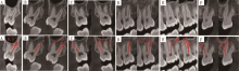
Fig.1
Curvature of the palatal root canal in BL and MD orientation"

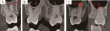
Fig.2
Distance and relationship between posterior roots and maxillary sinus floor"

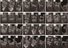
Fig.3
Several typical types of canal configuration in MSM with fused roots"

Tab.2
Types of root canal curvature of single canal maxillary second molars in BL and MD orientationsn n(%)"
| 根管 | 颊舌方向 | 近远中方向 | P | |||||
|---|---|---|---|---|---|---|---|---|
| Ⅰ型 | Ⅱ型 | Ⅲ型 | Ⅰ型 | Ⅱ型 | Ⅲ型 | |||
| 1R1C | 5(55.6) | 2(22.2) | 2(22.2) | 7(70.0) | 1(10.0) | 2(20.0) | 0.820 | |
| 2R2C | B(M) | 8(80.0) | 1(10.0) | 1(10.0) | 7(70.0) | 1(10.0) | 2(20.0) | 1.000 |
| P(D) | 2(20.0) | 6(60.0) | 2(20.0) | 8(80.0) | 2(20.0) | 0(0.0) | 0.033* | |
| 3R3C | MB | - | - | - | 1(0.4) | 22(8.9) | 225(90.7) | - |
| DB | 174(71.6) | 48(19.8) | 21(8.6) | 76(30.9) | 127(51.6) | 43(17.5) | 0.000* | |
| P | 99(39.8) | 97(38.9) | 53(21.3) | 205(82.7) | 32(12.9) | 11(4.4) | 0.000* | |
Tab.3
Relationship and distance between roots and the maxillary sinus floor"
| 牙根数 | 牙根位置 | 牙根的数目与出现频率(n(%)) | 合计 | 牙根到上颌窦底距离 | |||
|---|---|---|---|---|---|---|---|
| IS型 | CO型 | OS型 | 平均值/mm | 最小值至最大值/mm | |||
| 1个根 | 26(2.0) | 38(2.9) | 16(1.2) | 80(6.1) | -0.9±2.6 | -8.0~3.9 | |
| 2个根 | B(M) | 24(1.9) | 28(2.2) | 27(2.1) | 79(6.2) | 0.0±2.5 | -6.6~8.0 |
| P(D) | 12(0.9) | 26(2.0) | 42(3.2) | 80(6.1) | -0.9±2.3 | -4.9~6.4 | |
| 3个根 | MB | 130(10.0) | 127(9.8) | 96(7.4) | 353(27.2) | -0.6±2.6 | -7.8~10.5 |
| DB | 89(6.9) | 136(10.5) | 127(9.8) | 352(27.2) | -0.4±2.4 | -5.2~10.6 | |
| P | 69(5.3) | 119(9.2) | 165(12.7) | 353(27.2) | -0.8±2.8 | -6.4~10.6 | |
| 合计 | 350(27.0) | 474 (36.6) | 473 (36.4) | 1 297(100) | -0.2±2.6 | -8.0~10.6 | |
Tab.4
Distance between roots and the maxillary sinus floor according to patients’ age"
| 年龄组/岁 | 牙根数 | 牙根到上颌窦底距离/mm | P |
|---|---|---|---|
| 15~ | 206 | -0.9±2.2a | 0.000* |
| 25~ | 570 | -0.2±2.4a,b | |
| 35~ | 317 | 0.7±2.9b,c | |
| 45~ | 160 | 1.1±2.4c | |
| 55~ | 43 | 2.6±2.6d |
Tab.5
Number of root canals in maxillary permanent secondary molars reported in previous studies"
| 作者(年份) | 样本量 (n) | 地区 | 研究方法 | 1个根管/% | 2个根管/% | 3个根管/% | 4个根管/% | 5个根管/% |
|---|---|---|---|---|---|---|---|---|
| Olczak等(2017)[ | 207 | 波兰 | CBCT | 1.0 | 3.9 | 70.0 | 23.2 | - |
| 李建华(2017)[ | 607 | 中国山东 | CBCT | 2.31 | 7.41 | 72.32 | 17.96 | - |
| Kim等(2012)[ | 775 | 韩国 | CBCT | 3.9 | 8.1 | 56.9 | 30.8 | 0.3 |
| Tian等(2016)[ | 1539 | 中国上海 | CBCT | 3.2 | 6.1 | 66.4 | 22.9 | 1.4 |
| Xia等(2020)[ | 400 | 中国重庆 | CBCT | 1.25 | 11.25 | 61.75 | 25.75 | - |
| Felsypremila等(2015)[ | 371 | 印度 | CBCT | 2.4 | 7.3 | 38.8 | 50.4 | 0.5 |
| Ahmed Mostafa Ghobashy等(2017)[ | 610 | 埃及 | CBCT | 1.64 | 10.66 | 36.89 | 50.82 | - |
| Mirza等(2022)[ | 668 | 沙特阿拉伯 | CBCT | 0.6 | 1.5 | 53.3 | 44.3 | 0.3 |
| Silva等(2014)[ | 306 | 巴西 | CBCT | 1.96 | 7.84 | 55.88 | 34.31 | - |
| [1] | Kucher M, Dannemann M, Modler N, et al. Continuous measurement of three-dimensional root canal curvature using cone-beam computed and micro-computed tomography: A comparative study[J]. Dent J (Basel), 2020, 8(1): 16. |
| [2] |
Kang SH, Kim BS, Kim Y. Proximity of posterior teeth to the maxillary sinus and buccal bone thickness: A biometric assessment using cone-beam computed tomography[J]. J Endod, 2015, 41(11): 1839-1846.
doi: 10.1016/j.joen.2015.08.011 pmid: 26411520 |
| [3] |
Hauman CHJ, Chandler NP, Tong DC. Endodontic implications of the maxillary sinus: A review[J]. Int Endod J, 2002, 35(2): 127-141.
doi: 10.1046/j.0143-2885.2001.00524.x pmid: 11843967 |
| [4] | Ghasemi N, Rahimi S, Shahi S, et al. A review on root anatomy and canal configuration of the maxillary second molars[J]. Iran Endod J, 2017, 12(1): 1-9. |
| [5] | 李建华. 山东地区人群上颌第一、第二磨牙牙根及根管解剖结构的CBCT研究[D]. 济南: 山东大学, 2017. |
| [6] | Tian XM, Yang XW, Qian L, et al. Analysis of the root and canal morphologies in maxillary first and second molars in a Chinese population using cone-beam computed tomography[J]. J Endod, 2016, 42(5): 696-701. |
| [7] |
Kim Y, Lee SJ, Woo J. Morphology of maxillary first and second molars analyzed by cone-beam computed tomography in a Korean population: Variations in the number of roots and canals and the incidence of fusion[J]. J Endod, 2012, 38(8): 1063-1068.
doi: 10.1016/j.joen.2012.04.025 pmid: 22794206 |
| [8] |
Patel S, Wilson R, Dawood A, et al. The detection of periapical pathosis using periapical radiography and cone beam computed tomography-part 1: Pre-operative status[J]. Int Endod J, 2012, 45(8): 702-710.
doi: 10.1111/j.1365-2591.2011.01989.x pmid: 22188219 |
| [9] | 高静, 申静. 根尖周病中锥形束CT与根尖片识别病损差异的研究进展[J]. 华西口腔医学杂志, 2015, 33(2): 209-213. |
| [10] |
Venskutonis T, Plotino G, Juodzbalys G, et al. The importance of cone-beam computed tomography in the management of endodontic problems: A review of the literature[J]. J Endod, 2014, 40(12): 1895-1901.
doi: 10.1016/j.joen.2014.05.009 pmid: 25287321 |
| [11] | Oishi S, Ishida Y, Matsumura T, et al. A cone-beam computed tomographic assessment of the proximity of the maxillary canine and posterior teeth to the maxillary sinus floor: Lessons from 4778 roots[J]. Am J Orthod Dentofacial Orthop, 2020, 157(6): 792-802. |
| [12] | Tzeng LT, Chang MC, Chang SH, et al. Analysis of root canal system of maxillary first and second molars and their correlations by cone beam computed tomography[J]. J Formos Med Assoc, 2020, 119(5): 968-973. |
| [13] |
Zhang QY, Chen H, Fan B, et al. Root and root canal morphology in maxillary second molar with fused root from a native Chinese population[J]. J Endod, 2014, 40(6): 871-875.
doi: 10.1016/j.joen.2013.10.035 pmid: 24862720 |
| [14] | Schneider SW. A comparison of canal preparations in straight and curved root canals[J]. Oral Surg Oral Med Oral Pathol, 1971, 32(2): 271-275. |
| [15] | Tian XM, Qian L, Xin XZ, et al. An analysis of the proximity of maxillary posterior teeth to the maxillary sinus using cone-beam computed tomography[J]. J Endod, 2016, 42(3): 371-377. |
| [16] | Carrotte P. Endodontics: Part 8. Filling the root canal system[J]. Br Dent J, 2004, 197(11): 667-672. |
| [17] | Mirza MB, Gufran K, Alhabib O, et al. CBCT based study to analyze and classify root canal morphology of maxillary molars-A retrospective study[J]. Eur Rev Med Pharmacol Sci, 2022, 26(18): 6550-6560. |
| [18] |
Ratanajirasut R, Panichuttra A, Panmekiate S. A cone-beam computed tomographic study of root and canal morphology of maxillary first and second permanent molars in a Thai population[J]. J Endod, 2018, 44(1): 56-61.
doi: S0099-2399(17)30964-0 pmid: 29061352 |
| [19] |
Olczak K, Pawlicka H. The morphology of maxillary first and second molars analyzed by cone-beam computed tomography in a Polish population[J]. BMC Med Imaging, 2017, 17(1): 68.
doi: 10.1186/s12880-017-0243-3 pmid: 29284426 |
| [20] | Special Committee to Revise the Joint AAE/AAOMR Position Statement on use of CBCT in Endodontics. AAE and AAOMR joint position statement: Use of cone beam computed tomography in endodontics 2015 update[J]. Oral Surg Oral Med Oral Pathol Oral Radiol, 2015, 120(4): 508-512. |
| [21] |
American Dental Association Council on Scientific Affairs. The use of cone-beam computed tomography in dentistry: An advisory statement from the American Dental Association Council on Scientific Affairs[J]. J Am Dent Assoc, 2012, 143(8): 899-902.
doi: 10.14219/jada.archive.2012.0295 pmid: 22855905 |
| [22] | Xia Y, Qiao X, Huang YJ, et al. Root anatomy and root canal morphology of maxillary second permanent molars in a Chongqing population: A cone-beam computed tomography study[J]. Med Sci Monit, 2020, 26: e922794. |
| [23] |
Felsypremila G, Vinothkumar TS, Kandaswamy D. Anatomic symmetry of root and root canal morphology of posterior teeth in Indian subpopulation using cone beam computed tomography: A retrospective study[J]. Eur J Dent, 2015, 9(4): 500-507.
doi: 10.4103/1305-7456.172623 pmid: 26929687 |
| [24] |
Ghobashy AM, Nagy MM, Bayoumi AA. Evaluation of root and canal morphology of maxillary permanent molars in an Egyptian population by cone-beam computed tomography[J]. J Endod, 2017, 43(7): 1089-1092.
doi: S0099-2399(17)30259-5 pmid: 28476465 |
| [25] | Silva EJNL, Nejaim Y, Silva AIV, et al. Evaluation of root canal configuration of maxillary molars in a Brazilian population using cone-beam computed tomographic imaging: An in vivo study[J]. J Endod, 2014, 40(2): 173-176. |
| [26] |
Yang ZP, Yang SF, Lee G. The root and root canal anatomy of maxillary molars in a Chinese population[J]. Endod Dent Traumatol, 1988, 4(5): 215-218.
doi: 10.1111/j.1600-9657.1988.tb00324.x pmid: 3248579 |
| [27] |
Rwenyonyi CM, Kutesa AM, Muwazi LM, et al. Root and canal morphology of maxillary first and second permanent molar teeth in a Ugandan population[J]. Int Endod J, 2007, 40(9): 679-683.
doi: 10.1111/j.1365-2591.2007.01265.x pmid: 17608678 |
| [28] | Rouhani A, Bagherpour A, Akbari M, et al. Cone-beam computed tomography evaluation of maxillary first and second molars in Iranian population: A morphological study[J]. Iran Endod J, 2014, 9(3): 190-194. |
| [29] |
Ordinola-Zapata R, Martins JNR, Bramante CM, et al. Morphological evaluation of maxillary second molars with fused roots: A micro-CT study[J]. Int Endod J, 2017, 50(12): 1192-1200.
doi: 10.1111/iej.12752 pmid: 28196285 |
| [30] | Qiao X, Xu TT, Chen L, et al. Analysis of root canal curvature and root canal morphology of maxillary posterior teeth in Guizhou, China[J]. Med Sci Monit, 2021, 27: e928758. |
| [31] |
Jang JK, Kwak SW, Ha JH, et al. Anatomical relationship of maxillary posterior teeth with the sinus floor and buccal cortex[J]. J Oral Rehabil, 2017, 44(8): 617-625.
doi: 10.1111/joor.12525 pmid: 28547776 |
| [32] |
Ok E, Güngör E, Colak M, et al. Evaluation of the relationship between the maxillary posterior teeth and the sinus floor using cone-beam computed tomography[J]. Surg Radiol Anat, 2014, 36(9): 907-914.
doi: 10.1007/s00276-014-1317-3 pmid: 24874032 |
| [33] |
Zhang X, Li Y, Zhang Y, et al. Investigating the anatomical relationship between the maxillary molars and the sinus floor in a Chinese population using cone-beam computed tomography[J]. BMC Oral Health, 2019, 19(1): 282.
doi: 10.1186/s12903-019-0969-0 pmid: 31842859 |
| [34] | Jung YH, Cho BH. Assessment of the relationship between the maxillary molars and adjacent structures using cone beam computed tomography[J]. Imaging Sci Dent, 2012, 42(4): 219-224. |
| Viewed | ||||||
|
Full text |
|
|||||
|
Abstract |
|
|||||