
Stomatology ›› 2026, Vol. 46 ›› Issue (10): 771-775.doi: 10.13591/j.cnki.kqyx.2026.10.008
• Basic and Clinical Research • Previous Articles Next Articles
DING Yu1, GU Yongjia1( ), ZOU Derong2, YU Lüfeng2
Received:2025-10-08
Online:2026-10-28
Published:2026-10-16
CLC Number:
DING Yu, GU Yongjia, ZOU Derong, YU Lüfeng. Study on the condyle position in Class Ⅱ malocclusion patients with clinical symptoms of temporomandibular joint disorder[J]. Stomatology, 2026, 46(10): 771-775.
Fig.1
Measurement of the condyle volume"
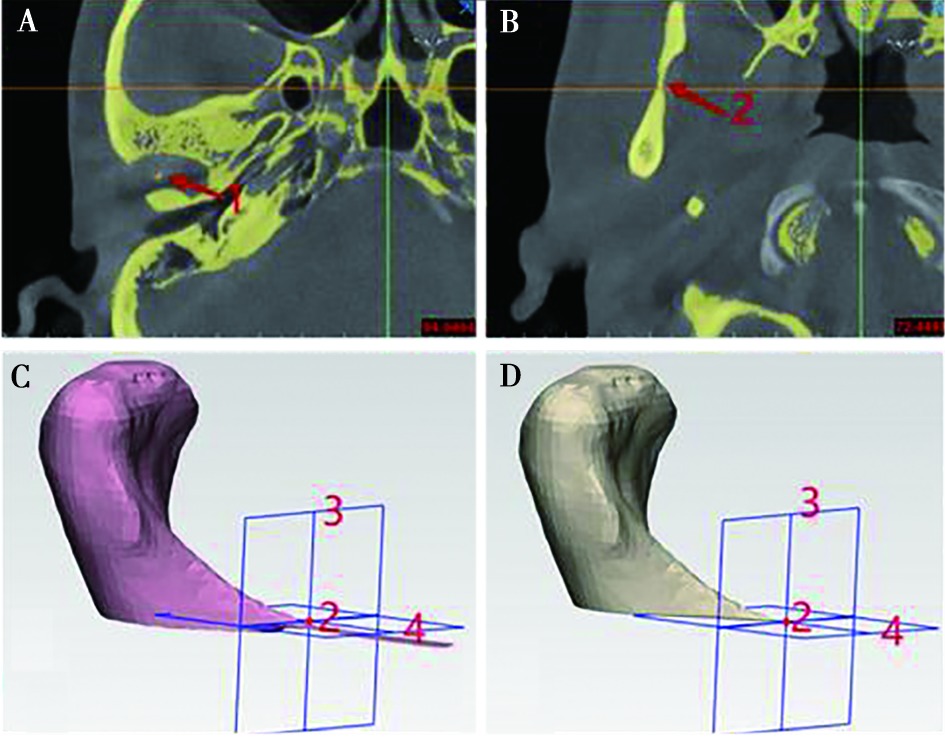
Fig.2
Measurement of the volume of the joint fossa"
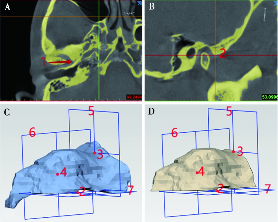
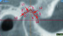
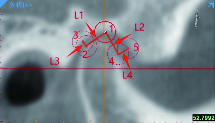
Fig.3
Measurement of the anterior joint space and the posterior joint space"
Tab.1
Comparison of measurement indexes between the NTMD group and TMD group"
| 指标 | 侧别 | NTMD组 | TMD组 | 组间 | 双侧比较 | |||||||
|---|---|---|---|---|---|---|---|---|---|---|---|---|
| NTMD组 | TMD组 | |||||||||||
| 年龄/岁 | 28.83±5.98 | 28.86±6.74 | 0.885 | |||||||||
| 髁突体积/mm3 | 右 | 2 302.71±546.35 | 2 299.19±810.53 | 0.983 | <0.001 | <0.001 | ||||||
| 左 | 1 679.06±356.07 | 1 683.93±519.61 | 0.964 | |||||||||
| 关节窝体积/mm3 | 右 | 1 034.19±285.26 | 1 097.11±314.54 | 0.307# | 0.654 | 0.457 | ||||||
| 左 | 1 022.29±296.03 | 1 116.36±291.74 | 0.104# | |||||||||
| C-FV | 右 | 2.33±0.68 | 2.17±0.68 | 0.323 | 0.833 | 0.535 | ||||||
| 左 | 2.35±0.63 | 2.12±0.71 | 0.165 | |||||||||
| 髁突水平角/(°) | 右 | 13.00±6.26 | 13.41±7.31 | 0.801# | 0.486 | 0.015 | ||||||
| 左 | 13.47±5.49 | 11.30±6.56 | 0.147# | |||||||||
| PJS/mm | 右 | 2.19±0.70 | 2.14±0.82 | 0.270# | 0.664 | 0.700& | ||||||
| 左 | 2.24±0.77 | 2.12±0.64 | 0.719# | |||||||||
| AJS/mm | 右 | 2.03±0.69 | 2.20±0.69 | 0.313# | 0.677 | 0.517& | ||||||
| 左 | 2.06±0.57 | 2.16±0.62 | 0.468# | |||||||||
Tab.2
Comparison of sagittal condyle position between the NTMD group and TMD group"
| 侧别 | 组别 | 髁突矢状向位置/n(%) | 卡方检验 | ||||||||
|---|---|---|---|---|---|---|---|---|---|---|---|
| 后位 | 中位 | 前位 | χ2 | P | |||||||
| 右 | NTMD组 | 4(13.3%) | 16(53.3%) | 10(33.3%) | 4.503 | 0.105 | |||||
| TMD组 | 10(27.0%) | 22(59.5%) | 5(13.5%) | ||||||||
| 左 | NTMD组 | 6(20.0%) | 17(56.7%) | 7(23.3%) | 1.313 | 0.519 | |||||
| TMD组 | 11(29.7%) | 16(43.2%) | 10(27.0%) | ||||||||
| [1] |
Edwards DC, Bowes CC, Penlington C, et al. Temporomandibular disorders and dietary changes: A cross-sectional survey[J]. J Oral Rehabil, 2021, 48(8): 873-879.
doi: 10.1111/joor.v48.8 |
| [2] |
Choudhary A, Ahuja U, Rathore A, et al. Association of temporomandibular joint morphology in patients with and without temporomandibular joint dysfunction: A cone-beam computed tomography based study[J]. Dent Res J, 2020, 17(5): 338.
doi: 10.4103/1735-3327.294334 |
| [3] | 王美青. 口腔解剖生理学[M]. 7版. 北京: 人民卫生出版社,2012:111. |
| [4] |
Bae JH, Hwang JY, Lee JY, et al. Change of TMJ space volume after mandibular setback BSSRO with intended manual condyle positioning in severe facial asymmetric patients[J]. J Craniofacial Surg, 2025, 36(4): e446-e449.
doi: 10.1097/SCS.0000000000011026 |
| [5] |
Noh KJ, Baik HS, Han S-S, et al. Differences in mandibular condyle and glenoid Fossa morphology in relation to vertical and sagittal skeletal patterns: A cone-beam computed tomography study[J]. Korean J Orthod, 2021, 51(2): 126-134.
doi: 10.4041/kjod.2021.51.2.126 pmid: 33678628 |
| [6] | 赵宁, 房兵. 正畸治疗与颞下颌关节紊乱病的研究进展[J]. 口腔医学, 2024, 44(1):20-23. |
| [7] | 杨乾. 口腔正畸患者颞下颌关节骨关节病的临床分析[J]. 中国实用医药, 2022, 17(17):73-75. |
| [8] |
Al-Rawi NH, Uthman AT, Sodeify SM. Spatial analysis of mandibular condyles in patients with temporomandibular disorders and normal controls using cone beam computed tomography[J]. Eur J Dent, 2017, 11(1): 99-105.
doi: 10.4103/ejd.ejd_202_16 pmid: 28435374 |
| [9] |
Derwich M, Pawlowska E. Do the mandibular condyles change their positions within glenoid fossae after occlusal splint therapy combined with physiotherapy in patients diagnosed with temporomandibular joint disorders? A prospective case control study[J]. J Pers Med, 2022, 12(2): 254.
doi: 10.3390/jpm12020254 |
| [10] |
Tǎut M, Barbur I, Hedeᶊiu M, et al. Condylar remodeling and skeletal changes following occlusal splint and manual therapy: A cone beam computed tomography study in temporomandibular disorders[J]. J Clin Med, 2024, 13(18): 5567.
doi: 10.3390/jcm13185567 |
| [11] |
Kadekuzhi S, Karuveettil V, Prabha RD, et al. Morphological changes in temporomandibular joint architecture in patients with temporomandibular disorders: Systematic review protocol[J]. BMJ Open, 2024, 14(9): e082396.
doi: 10.1136/bmjopen-2023-082396 |
| [12] |
De Carli E, Lagou A, Kiliaridis S, et al. Mandibular condyle changes in rats with unilateral masticatory function[J]. Orthod Craniofacial Res, 2023, 26(1): 37-45.
doi: 10.1111/ocr.v26.1 |
| [13] |
Zhou J, Yang HF, Li Q, et al. Comparison of temporomandibular joints in relation to ages and vertical facial types in skeletal Class Ⅱ female patients: A multiple-cross-sectional study[J]. BMC Oral Health, 2024, 24(1): 467.
doi: 10.1186/s12903-024-04219-4 |
| [14] |
Ren L, Chen PY, Musa MZ, et al. Quantitative and qualitative condylar changes Post-Stabilization splint in patients with temporomandibular disorder and chewing side preference[J]. Sci Rep, 2025, 15: 10996.
doi: 10.1038/s41598-025-94746-z |
| [15] |
Rovira-Lastra B, Flores-Orozco EI, Ayuso-Montero R, et al. Peripheral, functional and postural asymmetries related to the preferred chewing side in adults with natural dentition[J]. J Oral Rehabil, 2016, 43(4): 279-285.
doi: 10.1111/joor.12369 pmid: 26549578 |
| [16] |
Haralur SB, Majeed MI, Chaturvedi S, et al. Association between preferred chewing side and dynamic occlusal parameters[J]. J Int Med Res, 2019, 47(5): 1908-1915.
doi: 10.1177/0300060519827165 pmid: 30764682 |
| [17] |
Wojciechowska B, Szarmach A, Michcik A, et al. Association between clinical manifestations in temporomandibular joint disorders and corresponding radiographic findings[J]. J Clin Med, 2024, 13(16): 4886.
doi: 10.3390/jcm13164886 |
| [18] |
Shao B, Li C-X, Liu X, et al. Investigation of morphologic changes in temporomandibular joint with anterior disk displacement based on 3-dimensional reconstructive imaging[J]. J Craniofacial Surg, 2023, 34(4): 1271-1277.
doi: 10.1097/SCS.0000000000009139 |
| [19] | Krisjane Z, Urtane I, Krumina G, et al. Three-dimensional evaluation of TMJ parameters in Class Ⅱ and Class Ⅲ patients[J]. Stomatologija, 2009, 11(1): 32-36. |
| [20] | 刘旭, 李晨曦, 龚忠诚, 等. 关节盘前移位对颞下颌关节骨性结构关系影响的三维影像学测量研究[J]. 中华口腔医学杂志, 2024, 59(2):157-164. |
| [21] |
Alhammadi MS, Almashraqi AA, Thawaba AA, et al. Dimensional and positional temporomandibular joint osseous characteristics in normodivergent facial patterns with and without temporomandibular disorders[J]. Clin Oral Investig, 2023, 27(9): 5011-5020.
doi: 10.1007/s00784-023-05120-0 |
| [22] |
Almashraqi AA, Aboalnaga AA, Alhammadi MS, et al. Positional and dimensional osseous characteristics of the temporomandibular joint in female patients with skeletal Class Ⅲ malocclusion and disc displacement, with and without reduction[J]. Front Oral Health, 2025, 6: 1572305.
doi: 10.3389/froh.2025.1572305 |
| [23] | 谷志远, 傅开元, 张正康. 颞下颌关节紊乱病[M]. 北京: 人民卫生出版社,2008:111. |
| [24] |
Aboalnaga AA, Amer NM, Alhammadi MS, et al. Positional and dimensional TMJ characteristics in different temporomandibular disorders: A cross-sectional comparative study[J]. Cranio, 2024, 42(5): 611-619.
doi: 10.1080/08869634.2022.2028115 |
| [25] |
Zhang Z, Luo X, Li XQ, et al. The preliminary study of the effects of individual musculoskeletally stable position in the treatment of temporomandibular disorders[J]. BMC Oral Health, 2024, 24(1): 1083.
doi: 10.1186/s12903-024-04864-9 pmid: 39272167 |
| [26] | 樊文萍, 刘梦琦, 张晓欢. 颞下颌关节紊乱病患者髁突位置和形态的MRI观察[J]. 中华口腔医学杂志, 2019, 58(8):522-526. |
| [27] | Shahab S, Amoozad Khalili Z, Emami Meybodi E, et al. Relation between condyle horizontal angle and intercondylar angle with disc displacement in patients with temporomandibular joint disorders: An MRI evaluation[J]. Radiol Res Pract, 2023, 2023: 3846525. |
| [28] |
Gao YD, Luo D, Yuan MJ, et al. The relationship between the oblique sagittal temporomandibular joint disc position and the volume surface area of the condyle in young TMD adults[J]. Front Bioeng Biotechnol, 2023, 11: 1321241.
doi: 10.3389/fbioe.2023.1321241 |
| Viewed | ||||||
|
Full text |
|
|||||
|
Abstract |
|
|||||
