
Stomatology ›› 2025, Vol. 45 ›› Issue (6): 445-452.doi: 10.13591/j.cnki.kqyx.2025.06.009
• Basic and Clinical Research • Previous Articles Next Articles
LIU Fei1,2,3, ZHANG Jiulou4( ), JIN Ruofan1,2,3, ZHANG Nan1,2,3, ZHOU Weina1,2,3()
Received:2024-12-29
Online:2025-06-28
Published:2025-07-08
CLC Number:
LIU Fei, ZHANG Jiulou, JIN Ruofan, ZHANG Nan, ZHOU Weina. The automatic segmentation of the temporomandibular joint based on MRI using deep learning method[J]. Stomatology, 2025, 45(6): 445-452.
Tab.1
MRI findings of all subjects"
| 关节盘位置 | 左侧 | 右侧 |
|---|---|---|
| 正常 | 43 | 39 |
| ADDwR | 25 | 23 |
| ADDwoR | 36 | 42 |
| 总计 | 104 | 104 |
Tab.2
MRI findings in the test(validation)set"
| 关节盘位置 | 左侧 | 右侧 |
|---|---|---|
| 正常 | 6 | 3 |
| ADDwR | 6 | 6 |
| ADDwoR | 8 | 11 |
| 总计 | 20 | 20 |

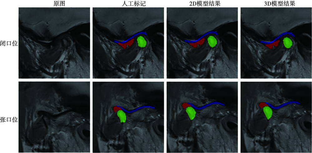
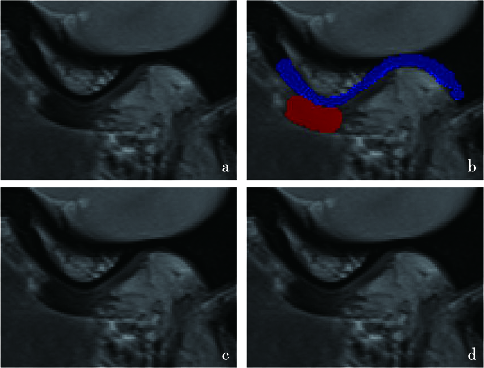
Fig.1
Example of raw image(a)and labeled image(b), where the red part is the articular disc,the green part is the condyle,and the blue part is the glenoid fossa"
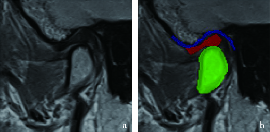

Fig.2
Flow diagram of 2D model"
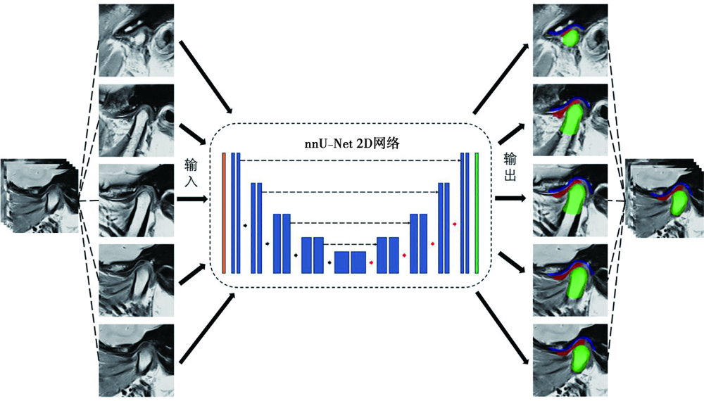
Fig.3
Flow diagram of 3D model"
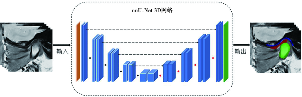
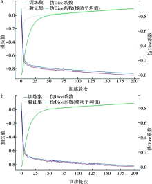
Fig.4
Convergence curve of nnU-Net model training"
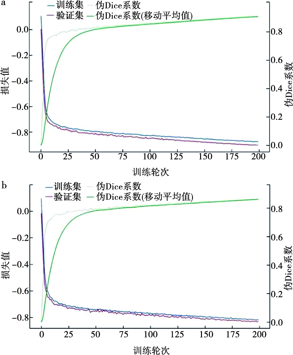
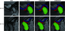
Fig.5
MRI image without ADD"

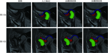
Fig.6
MRI image with ADDwR"
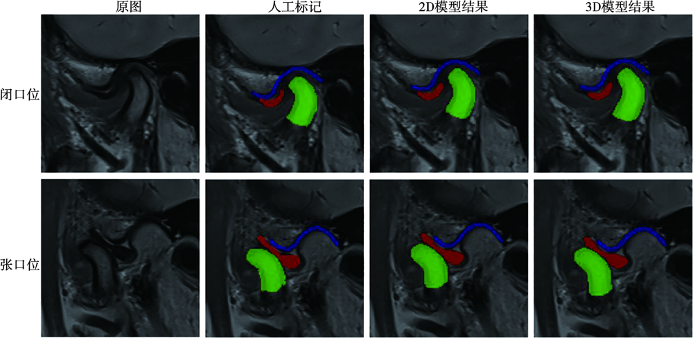
Fig.7
MRI image with ADDwoR"
Tab.3
The Dice, Recall, Precision and F1 score of 2D/3D model in the close or open mouth images"
| 模型 | 图像类别 | 关节组织 | Dice | 召回率 | 精确率 | F1分数 |
|---|---|---|---|---|---|---|
| 2D模型 | 闭口位 | 关节盘 | 0.775 911 | 0.761 949 | 0.802 372 | 0.781 638 |
| 髁突 | 0.861 552 | 0.858 067 | 0.880 847 | 0.869 308 | ||
| 关节窝 | 0.661 321 | 0.613 758 | 0.717 471 | 0.661 574 | ||
| 开口位 | 关节盘 | 0.790 665 | 0.788 616 | 0.807 662 | 0.798 026 | |
| 髁突 | 0.899 456 | 0.901 339 | 0.908 778 | 0.905 043 | ||
| 关节窝 | 0.662 487 | 0.624 911 | 0.711 179 | 0.665 260 | ||
| 3D模型 | 闭口位 | 关节盘 | 0.778 488 | 0.748 471 | 0.821 614 | 0.783 339 |
| 髁突 | 0.869 087 | 0.871 318 | 0.882 744 | 0.876 994 | ||
| 关节窝 | 0.680 833 | 0.643 324 | 0.722 766 | 0.680 735 | ||
| 开口位 | 关节盘 | 0.796 855 | 0.781 431 | 0.827 924 | 0.804 006 | |
| 髁突 | 0.902 941 | 0.906 160 | 0.912 518 | 0.909 328 | ||
| 关节窝 | 0.680 003 | 0.650 240 | 0.716 696 | 0.681 853 |
Tab.4
Results of One-way ANOVA test:there was no significant difference in the segmentation ability of the four models(2D/3D models in the open and closed images)"
| 关节组织 | 平方和 | 自由度 | 均方 | F | P | |
|---|---|---|---|---|---|---|
| 关节盘 | 组间 | 0.006 | 3 | 0.002 | 0.350 | 0.789 |
| 组内 | 0.429 | 76 | 0.006 | |||
| 总计 | 0.435 | 79 | ||||
| 髁突 | 组间 | 0.026 | 3 | 0.009 | 1.998 | 0.121 |
| 组内 | 0.335 | 76 | 0.004 | |||
| 总计 | 0.362 | 79 | ||||
| 关节窝 | 组间 | 0.007 | 3 | 0.002 | 0.376 | 0.771 |
| 组内 | 0.263 | 57 | 0.005 | |||
| 总计 | 0.471 | 79 |
Tab.5
Results of One-way ANOVA test:there were statistical differences in the segmentation effect of the three joint tissues(articular disc,condyle,and glenoid fossa)"
| 模型 | 平方和 | 自由度 | 均方 | F | P | |
|---|---|---|---|---|---|---|
| 2D/闭口位 | 组间 | 0.404 | 2 | 0.202 | 34.933 | <0.001 |
| 组内 | 0.329 | 57 | 0.006 | |||
| 总计 | 0.733 | 59 | ||||
| 3D/闭口位 | 组间 | 0.355 | 2 | 0.177 | 30.568 | <0.001 |
| 组内 | 0.331 | 57 | 0.006 | |||
| 总计 | 0.685 | 59 | ||||
| 2D/开口位 | 组间 | 0.563 | 2 | 0.281 | 52.560 | <0.001 |
| 组内 | 0.305 | 57 | 0.005 | |||
| 总计 | 0.868 | 59 | ||||
| 3D/开口位 | 组间 | 0.497 | 2 | 0.249 | 53.872 | <0.001 |
| 组内 | 0.263 | 57 | 0.005 | |||
| 总计 | 0.761 | 59 |
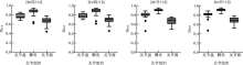
Fig.8
Plot of Dice distribution for each singleton of the test set,with each dot representing an outlier"

Tab.6
The diagnostic accuracy of ADD by young doctors based on the original MRI image and the segmentation results of 2D and 3D models(with the senior doctor’s diagnosis for the correct diagnosis) %"
| 诊断依据 | 正常 | ADDwR | ADDwoR | 总计 |
|---|---|---|---|---|
| 原图 | 66.7 | 83.3 | 88.9 | 82.5 |
| 2D网络分割图 | 88.9 | 100 | 100 | 97.5 |
| 2D网络分割图 | 88.9 | 100 | 100 | 97.5 |
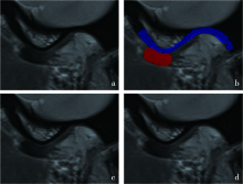
Fig.9
Dice coefficient discretization case example diagram"
| [1] | 谢丛蔓. 中国大陆地区颞下颌关节紊乱病流行趋势的系统评价及Meta分析[D]. 重庆: 重庆医科大学, 2020. |
| [2] | Lei J, Yap AU, Li Y, et al. Clinical protocol for managing acute disc displacement without reduction: A magnetic resonance imaging evaluation[J]. Int J Oral Maxillofac Surg, 2020, 49(3): 361-368. |
| [3] |
Spitzer WJ, Lenz M, Sauter R. Imaging the articular disk of the temporomandibular joint using nuclear magnetic resonance: Preliminary report[J]. Dtsch Zahnarztl Z, 1986, 41(7): 693-696.
pmid: 3461994 |
| [4] | 傅开元, 雷杰. 颞下颌关节紊乱病的分类、诊断及治疗进展[J]. 口腔医学, 2024, 44(1): 6-10. |
| [5] | 张倩, 李俊, 马丽霞, 等. 颞下颌关节张闭口位磁共振成像在颞下颌关节盘前移位中的诊断价值[J]. 影像研究与医学应用, 2021, 5(14): 26-28. |
| [6] | Widmalm SE, Brooks SL, Sano T, et al. Limitation of the diagnostic value of MR images for diagnosing temporomandibular joint disorders[J]. Dentomaxillofac Radiol, 2006, 35(5): 334-338. |
| [7] | Nebbe B, Brooks SL, Hatcher D, et al. Magnetic resonance imaging of the temporomandibular joint: Interobserver agreement in subjective classification of disk status[J]. Oral Surg Oral Med Oral Pathol Oral Radiol Endod, 2000, 90(1): 102-107. |
| [8] | Alkhader M, Ohbayashi N, Tetsumura A, et al. Diagnostic performance of magnetic resonance imaging for detecting osseous abnormalities of the temporomandibular joint and its correlation with cone beam computed tomography[J]. Dentomaxillofac Radiol, 2010, 39(5): 270-276. |
| [9] | LeCun Y, Bengio Y, Hinton G. Deep learning[J]. Nature, 2015, 521(7553): 436-444. |
| [10] | 殷晓航, 王永才, 李德英. 基于U-Net结构改进的医学影像分割技术综述[J]. 软件学报, 2021, 32(2): 519-550. |
| [11] | Savjani R. nnU-Net: Further automating biomedical image autosegmentation[J]. Radiol Imaging Cancer, 2021, 3(1): e209039. |
| [12] |
Isensee F, Jaeger PF, Kohl SAA, et al. nnU-Net: A self-configuring method for deep learning-based biomedical image segmentation[J]. Nat Methods, 2021, 18(2): 203-211.
doi: 10.1038/s41592-020-01008-z pmid: 33288961 |
| [13] | Zwijnen AW, Watzema L, Ridwan Y, et al. Self-adaptive deep learning-based segmentation for universal and functional clinical and preclinical CT image analysis[J]. Comput Biol Med, 2024, 179: 108853. |
| [14] | Kang H, Witanto JN, Pratama K, et al. Fully automated MRI segmentation and volumetric measurement of intracranial meningioma using deep learning[J]. J Magn Reson Imaging, 2023, 57(3): 871-881. |
| [15] | Bareja R, Ismail M, Martin D, et al. nnU-net-based segmentation of tumor subcompartments in pediatric medulloblastoma using multiparametric MRI: A multi-institutional study[J]. Radiol Artif Intell, 2024, 6(5): e230115. |
| [16] | 方媛媛, 毛伟玉, 王中振, 等. 基于深度学习的颞下颌关节骨关节病锥形束CT自动诊断及分类[C]// 中华口腔医学会颞下颌关节病学及牙合学专委会. 第20次全国颞下颌关节病学及牙合学研讨会暨第七届亚洲颞下颌关节学术大会论文汇编. 北京,2023:284-285. |
| [17] |
Ito S, Mine Y, Yoshimi Y, et al. Automated segmentation of articular disc of the temporomandibular joint on magnetic resonance images using deep learning[J]. Sci Rep, 2022, 12(1): 221.
doi: 10.1038/s41598-021-04354-w pmid: 34997167 |
| [18] | Lin BL, Cheng MS, Wang SZ, et al. Automatic detection of anteriorly displaced temporomandibular joint discs on magnetic resonance images using a deep learning algorithm[J]. Dentomaxillofac Radiol, 2022, 51(3): 20210341. |
| [19] | Nozawa M, Ito H, Ariji Y, et al. Automatic segmentation of the temporomandibular joint disc on magnetic resonance images using a deep learning technique[J]. Dentomaxillofac Radiol, 2022, 51(1): 20210185. |
| [20] | Emshoff R, Brandlmaier I, Bertram S, et al. Relative odds of temporomandibular joint pain as a function of magnetic resonance imaging findings of internal derangement, osteoarthrosis, effusion, and bone marrow edema[J]. Oral Surg Oral Med Oral Pathol Oral Radiol Endod, 2003, 95(4): 437-445. |
| [21] | Chan HP, Samala RK, Hadjiiski LM, et al. Deep learning in medical image analysis[J]. Adv Exp Med Biol, 2020, 1213: 3-21. |
| [22] | Li MX, Punithakumar K, Major PW, et al. Temporomandibular joint segmentation in MRI images using deep learning[J]. J Dent, 2022, 127: 104345. |
| [23] | Soldati E, Rossi F, Vicente J, et al. Survey of MRI usefulness for the clinical assessment of bone microstructure[J]. Int J Mol Sci, 2021, 22(5): 2509. |
| Viewed | ||||||
|
Full text |
|
|||||
|
Abstract |
|
|||||