
Stomatology ›› 2025, Vol. 45 ›› Issue (1): 58-63.doi: 10.13591/j.cnki.kqyx.2025.01.009
• Basic and Clinical Research • Previous Articles Next Articles
ZHU Peng1, GU Yongchun2( ), WU Yihan2, XU Xiaoming3
Received:2024-08-20
Online:2025-01-28
Published:2025-01-16
CLC Number:
ZHU Peng, GU Yongchun, WU Yihan, XU Xiaoming. The root canal morphology of mandibular anterior teeth and its correlation with the occurrence of three-rooted mandibular first molars[J]. Stomatology, 2025, 45(1): 58-63.
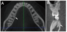
Fig.1
CBCT observation of the roots of mandibular first molars"


Fig.2
Mandibular anterior teeth with a single root and a single canal"
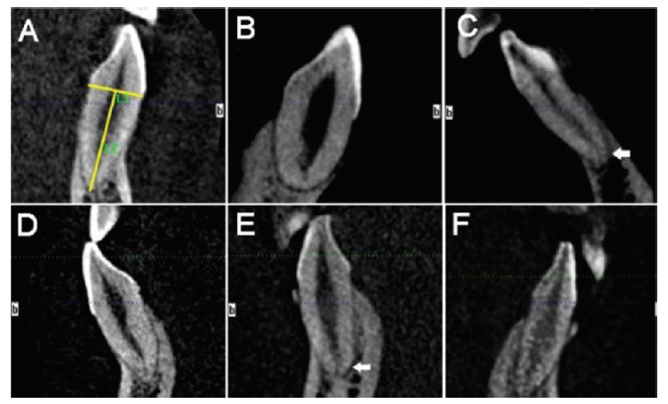

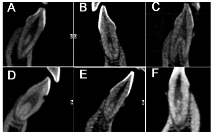
Fig.3
Mandibular anterior teeth with two root canals"
Tab.1
Measurement results of roots of mandibular anterior teeth mm"
| 项目 | 中切牙 | 侧切牙 | 尖牙 | |||
|---|---|---|---|---|---|---|
| 根长 | 牙颈部唇舌径 | 根长 | 牙颈部唇舌径 | 根长 | 牙颈部唇舌径 | |
| 左右侧 | ||||||
| 左侧 | 11.4±1.3 | 5.4±0.5 | 12.6±1.4 | 6.0±0.7 | 14.8±1.7 | 7.6±0.8 |
| 右侧 | 11.3±1.2 | 5.4±0.5 | 12.6±1.2 | 6.0±0.5 | 14.6±1.7 | 7.5±0.7 |
| 性别 | ||||||
| 男 | 11.7±1.3 | 5.6±0.4 | 12.8±1.3 | 6.1±0.5 | 15.1±1.9 | 7.9±0.8 |
| 女 | 11.0±1.0** | 5.3±0.4** | 12.3±1.2** | 5.9±0.7** | 14.3±1.4** | 7.2±0.5** |
Tab.2
Occurrence of two root canals in mandibular anterior teeth %"
| 项目 | 中切牙 | 侧切牙 | 尖牙 | |||
|---|---|---|---|---|---|---|
| 单根管 | 双根管 | 单根管 | 双根管 | 单根管 | 双根管 | |
| 左右侧 | ||||||
| 左侧 | 90.8 | 9.2 | 82.9 | 17.1 | 95.0 | 5.0 |
| 右侧 | 88.4 | 11.6 | 79.9 | 20.1 | 92.0 | 8.0 |
| 性别 | ||||||
| 男 | 89.9 | 10.1 | 79.9 | 20.1 | 95.5 | 4.5 |
| 女 | 89.2 | 10.8 | 82.9 | 17.1 | 91.5 | 8.5 |
| 合计 | 89.4 | 10.4 | 81.4 | 18.6 | 93.5 | 6.5 |
Tab.3
Types of root canal configurations in mandibular anterior teeth (Vertucci’s classification) n(%)"
| 根管类型 | 中切牙 | 侧切牙 | 尖牙 |
|---|---|---|---|
| 1-1 | 352(89.6) | 324(81.4) | 374(93.5) |
| 2-1 | 4(1.0) | 21(5.3) | 3(0.8) |
| 1-2-1 | 19(4.8) | 35(8.8) | 12(3.0) |
| 2-2 | 1(0.3) | 2(0.5) | 4(1.0) |
| 1-2 | 17(4.3) | 15(3.7) | 7(1.7) |
| 2-1-2 | 0(0) | 1(0.3) | 0(0) |
| 合计 | 393(100.0) | 398(100.0) | 400(100.0) |
Tab.4
Correlations between the bilateral antimetric mandibular anterior teeth with two root canals"
| 项目 | 下颌中切牙 | 下颌侧切牙 | 下颌尖牙 | |||||||
|---|---|---|---|---|---|---|---|---|---|---|
| 左侧双根管 | 左侧单根管 | 合计 | 左侧双根管 | 左侧单根管 | 合计 | 左侧双根管 | 左侧单根管 | 合计 | ||
| 右侧双根管 | 11 | 12 | 23 | 22 | 18 | 40 | 6 | 10 | 16 | |
| 右侧单根管 | 7 | 163 | 170 | 12 | 146 | 158 | 4 | 180 | 184 | |
| 合计 | 18 | 175 | 193 | 34 | 164 | 198 | 10 | 190 | 200 | |
| rho | 0.487 | 0.505 | 0.440 | |||||||
| P | 0.000 | 0.000 | 0.000 | |||||||
Tab.5
Bilateral symmetry of mandibular first molars’ root numbers"
| 下颌第一磨牙 | 男性 | 女性 | 合计 | ||||||||
|---|---|---|---|---|---|---|---|---|---|---|---|
| 左侧三根 | 左侧双根 | 合计 | 左侧三根 | 左侧双根 | 合计 | 左侧三根 | 左侧双根 | 合计 | |||
| 右侧三根 | 15 | 10 | 25 | 16 | 9 | 25 | 31 | 19 | 50 | ||
| 右侧双根 | 2 | 67 | 69 | 2 | 57 | 59 | 4 | 124 | 128 | ||
| 合计 | 17 | 77 | 94 | 18 | 66 | 84 | 35 | 143 | 178 | ||
| rho | 0.655 | 0.675 | 0.666 | ||||||||
| P | 0.000 | 0.000 | 0.000 | ||||||||
Tab.6
Correlations between the three-rooted mandibular first molar and double-canaled mandibular incisor on the left side"
| 下颌第 一磨牙 | 中切牙 | 侧切牙 | 尖牙 | ||||||
|---|---|---|---|---|---|---|---|---|---|
| 双根管 | 单根管 | 合计 | 双根管 | 单根管 | 合计 | 双根管 | 单根管 | 合计 | |
| 三根 | 5 | 33 | 38 | 5 | 34 | 39 | 7 | 32 | 39 |
| 双根 | 12 | 137 | 149 | 27 | 125 | 152 | 3 | 150 | 153 |
| 合计 | 17 | 170 | 187 | 32 | 159 | 191 | 10 | 182 | 192 |
| rho | 0.063 | 0.053 | 0.289 | ||||||
| P | 0.404 | 0.464 | 0.000 | ||||||
Tab.7
Correlations between the three-rooted mandibular first molar and double-canaled mandibular central/lateral incisor on the right side"
| 下颌第 一磨牙 | 中切牙 | 侧切牙 | 尖牙 | ||||||
|---|---|---|---|---|---|---|---|---|---|
| 双根管 | 单根管 | 合计 | 双根管 | 单根管 | 合计 | 双根管 | 单根管 | 合计 | |
| 三根 | 6 | 45 | 51 | 14 | 40 | 54 | 8 | 46 | 54 |
| 双根 | 13 | 119 | 132 | 21 | 110 | 131 | 8 | 124 | 132 |
| 合计 | 19 | 164 | 183 | 35 | 150 | 185 | 16 | 170 | 186 |
| rho | 0.028 | 0.115 | 0.142 | ||||||
| P | 0.705 | 0.119 | 0.054 | ||||||
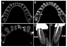
Fig.4
Bilateral symmetry and concurrence of double-canaled mandibular anterior incisors and three-rooted mandibular first molars"

| [1] |
Vertucci FJ. Root canal anatomy of the mandibular anterior teeth[J]. J Am Dent Assoc, 1974, 89(2): 369-371.
doi: 10.14219/jada.archive.1974.0391 pmid: 4527223 |
| [2] | Chen M, Wang H, Tsauo C, et al. Micro-computed tomography analysis of root canal morphology and thickness of crown and root of mandibular incisors in Chinese population[J]. Clin Oral Investig, 2022, 26(1): 901-910. |
| [3] |
Baxter S, Jablonski M, Hülsmann M. Cone-beam-computed-tomography of the symmetry of root canal anatomy in mandibular incisors[J]. J Oral Sci, 2020, 62(2): 180-183.
doi: 10.2334/josnusd.19-0113 pmid: 32224571 |
| [4] |
Han T, Ma Y, Yang L, et al. A study of the root canal morphology of mandibular anterior teeth using cone-beam computed tomography in a Chinese subpopulation[J]. J Endod, 2014, 40(9): 1309-1314.
doi: 10.1016/j.joen.2014.05.008 pmid: 25043332 |
| [5] | Jiang CF, Pei F, Wu YH, et al. Investigation of three-rooted deciduous mandibular second molars in a Chinese population using cone-beam computed tomography[J]. BMC Oral Health, 2022, 22(1): 329. |
| [6] | Qiao X, Zhu HL, Yan YJ, et al. Prevalence of middle mesial canal and Radix entomolaris of mandibular first permanent molars in a western Chinese population: An in vivo cone-beam computed tomographic study[J]. BMC Oral Health, 2020, 20(1): 224. |
| [7] | Gu YC, Lu Q, Wang HG, et al. Root canal morphology of permanent three-rooted mandibular first molars: Part Ⅰ-Pulp floor and root canal system[J]. J Endod, 2010, 36(6): 990-994. |
| [8] | 顾永春, 倪龙兴. X线投照角度对诊断下颌第一恒磨牙远舌根变异的作用[J]. 实用口腔医学杂志, 2013, 29(1): 94-96. |
| [9] | Tredoux S, Warren N, Buchanan GD. Root and canal configura-tions of mandibular first molars in a South African subpopulation[J]. J Oral Sci, 2021, 63(3): 252-256. |
| [10] | Kantilieraki E, Delantoni A, Angelopoulos C, et al. Evaluation of root and root canal morphology of mandibular first and second molars in a Greek population: A CBCT study[J]. Eur Endod J, 2019, 4(2): 62-68. |
| [11] |
Shigefuji R, Serikawa M, Usami A. Observation of mandibular second molar roots and root canal morphology using dental cone-beam computed tomography[J]. Anat Cell Biol, 2022, 55(2): 155-160.
doi: 10.5115/acb.22.050 pmid: 35773218 |
| [12] |
Martins JNR, Gu YC, Marques D, et al. Differences on the root and root canal morphologies between Asian and white ethnic groups analyzed by cone-beam computed tomography[J]. J Endod, 2018, 44(7): 1096-1104.
doi: S0099-2399(18)30239-5 pmid: 29861062 |
| [13] | Wu YC, Cheng WC, Chung MP, et al. Complicated root canal morphology of mandibular lateral incisors is associated with the presence of distolingual root in mandibular first molars: A cone-beam computed tomographic study in a Taiwanese population[J]. J Endod, 2018, 44(1): 73-79.e1. |
| [14] | Wu YC, Cheng WC, Weng PW, et al. The presence of distolingual root in mandibular first molars is correlated with complicated root canal morphology of mandibular central incisors: A cone-beam computed tomographic study in a Taiwanese population[J]. J Endod, 2018, 44(5): 711-716.e1. |
| [15] | Yeung AWK, Jacobs R, Bornstein MM. Novel low-dose protocols using cone beam computed tomography in dental medicine: A review focusing on indications, limitations, and future possibilities[J]. Clin Oral Investig, 2019, 23(6): 2573-2581. |
| [16] |
Patel S, Brown J, Pimentel T, et al. Cone beam computed tomography in Endodontics—A review of the literature[J]. Int Endod J, 2019, 52(8): 1138-1152.
doi: 10.1111/iej.13115 pmid: 30868610 |
| [17] | Bai BB, Tang Y, Wu YH, et al. Ex vivo detection of mandibular incisors’ root canal morphology using cone-beam computed tomography with 4 different voxel sizes and micro-computed tomography[J]. BMC Oral Health, 2023, 23(1): 656. |
| [18] |
Lin ZT, Hu QG, Wang TM, et al. Use of CBCT to investigate the root canal morphology of mandibular incisors[J]. Surg Radiol Anat, 2014, 36(9): 877-882.
doi: 10.1007/s00276-014-1267-9 pmid: 24515289 |
| [19] |
Liu J, Luo J, Dou L, et al. CBCT study of root and canal morphology of permanent mandibular incisors in a Chinese population[J]. Acta Odontol Scand, 2014, 72(1): 26-30.
doi: 10.3109/00016357.2013.775337 pmid: 24255962 |
| [20] | Yang ZY, Lu KK, Wang F, et al. Cone-beam computed tomography study of the root and canal morphology of mandibular permanent anterior teeth in a Chongqing population[J]. Ther Clin Risk Manag, 2015, 12: 19-25. |
| [21] | 赵莹, 董颖韬, 王晓燕, 等. 4674颗下颌前牙根管构型的锥形束CT分析[J]. 北京大学学报(医学版), 2014, 46(1): 95-99. |
| [22] |
Candeiro GTM, Monteiro Dodt Teixeira IM, Olimpio Barbosa DA, et al. Vertucci’s root canal configuration of 14 413 mandibular anterior teeth in a Brazilian population: A prevalence study using cone-beam computed tomography[J]. J Endod, 2021, 47(3): 404-408.
doi: 10.1016/j.joen.2020.12.001 pmid: 33326836 |
| [23] |
Mahmood TR. Assessment of root canal morphology of mandibular permanent anterior teeth in an Iraqi subpopulation by cone-beam computed tomography[J]. J Dent Sci, 2021, 16(4): 1182-1190.
doi: 10.1016/j.jds.2021.02.010 pmid: 34484586 |
| [24] |
Kabak YS, Abbott PV. Endodontic treatment of mandibular incisors with two root canals: Report of two cases[J]. Aust Endod J, 2007, 33(1): 27-31.
doi: 10.1111/j.1747-4477.2007.00054.x pmid: 17461838 |
| [25] | Gu YC, Lu Q, Wang P, et al. Root canal morphology of permanent three-rooted mandibular first molars: Part Ⅱ-Measurement of root canal curvatures[J]. J Endod, 2010, 36(8): 1341-1346. |
| [26] | Ishii N, Sakuma A, Makino Y, et al. Incidence of three-rooted mandibular first molars among contemporary Japanese individuals determined using multidetector computed tomography[J]. Leg Med, 2016, 22: 9-12. |
| [27] | Jang JK, Peters OA, Lee W, et al. Incidence of three roots and/or four root canals in the permanent mandibular first molars in a Korean sub-population[J]. Clin Oral Investig, 2013, 17(1): 105-111. |
| Viewed | ||||||
|
Full text |
|
|||||
|
Abstract |
|
|||||