
Stomatology ›› 2024, Vol. 44 ›› Issue (6): 469-474.doi: 10.13591/j.cnki.kqyx.2024.06.013
• Review • Previous Articles Next Articles
DU Xingping1,NI Peng1,ZHANG Lei2( )
)
Received:2023-07-10
Online:2024-06-28
Published:2024-06-27
CLC Number:
DU Xingping, NI Peng, ZHANG Lei. Cone beam CT study on the anatomical structure of the mandibular interforaminal area and its clinical significance[J]. Stomatology, 2024, 44(6): 469-474.
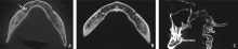
Fig.1
Sketch map of anterior loop (arrow showed)"

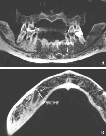
Fig.2
Sketch map of mandibular incisive canal"

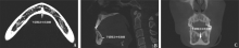
Fig.3
Sketch map of mandibular median lingual canal"

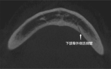
Fig.4
Sketch map of mandibular lateral lingual canal"

| [1] |
Goller Bulut D, Köse E. Availablebone morphology and status of neural structures in the mandibular interforaminal region: Three-dimensional analysis of anatomical structures[J]. Surg Radiol Anat, 2018, 40(11):1243-1252.
doi: 10.1007/s00276-018-2039-8 pmid: 29766231 |
| [2] | Puri A, Verma P, Mahajan P, et al. CBCT evaluation of the vitalmandibular interforaminal anatomical structures[J]. Ann Maxillofac Surg, 2020, 10(1):149-157. |
| [3] |
Caughey JA, Do Q, Shen D, et al. Comprehensive review of the incisive branch of the inferior alveolar nerve[J]. Anat Cell Biol, 2021, 54(4):409-416.
doi: 10.5115/acb.21.113 pmid: 34620736 |
| [4] |
于鸿滨, 钱石兵, 刘超峰, 等. 下颌正中舌侧管、下颌舌侧副管和下颌切牙神经管解剖关系的CBCT影像研究[J]. 口腔医学研究, 2023, 39(5):440-444.
doi: 10.13701/j.cnki.kqyxyj.2023.05.012 |
| [5] | Ferreira Barbosa DA, Barros ID, Teixeira RC, et al. Imaging aspects of the mandibular incisive canal: A PROSPERO-registered systematic review and meta-analysis of cone beam computed tomography studies[J]. Int J Oral Maxillofac Implants, 2019, 34(2):423-433. |
| [6] |
Krishnan U, Monsour P, Thaha K, et al. A limited field cone-beam computed tomography-based evaluation of the mental foramen, accessory mental foramina, anterior loop, lateral lingual foramen, and lateral lingual canal[J]. J Endod, 2018, 44(6):946-951.
doi: S0099-2399(18)30067-0 pmid: 29550007 |
| [7] |
Wei XY, Gu PM, Hao YS, et al. Detection and characterization of anterior loop, accessory mental foramen, and lateral lingual foramen by using cone beam computed tomography[J]. J Prosthet Dent, 2020, 124(3):365-371.
doi: S0022-3913(19)30494-9 pmid: 31787268 |
| [8] |
Liang X, Jacobs R, Lambrichts I, et al. Lingual foramina on the mandibular midline revisited: A macroanatomical study[J]. Clin Anat, 2007, 20(3):246-251.
doi: 10.1002/ca.20357 pmid: 16683247 |
| [9] |
He XJ, Jiang JQ, Cai W, et al. Assessment of the appearance, location and morphology of mandibular lingual foramina using cone beam computed tomography[J]. Int Dent J, 2016, 66(5):272-279.
doi: 10.1111/idj.12242 pmid: 27177988 |
| [10] | Yang XW, Zhang FF, Li YH, et al. Characteristics of intrabony nerve canals in mandibular interforaminal region by using cone-beam computed tomography and a recommendation of safe zone for implant and bone harvesting[J]. Clin Implant Dent Relat Res, 2017, 19(3):530-538. |
| [11] | Parnia F, Moslehifard E, Hafezeqoran A, et al. Characteristics of anatomicallandmarks in the mandibular interforaminal region: A cone-beam computed tomography study[J]. Med Oral Patol Oral Cir Bucal, 2012, 17(3):e420-e425. |
| [12] |
do Carmo Oliveira M, Tedesco TK, Gimenez T, et al. Analysis of the frequency of visualization of morphological variations in anatomical bone features in the mandibular interforaminal region through cone-beam computed tomography[J]. Surg Radiol Anat, 2018, 40(10):1119-1131.
doi: 10.1007/s00276-018-2040-2 pmid: 29785677 |
| [13] | Barbosa DA, Kurita LM, Pimenta AV, et al. Mandibular incisive canal-related prevalence, morphometric parameters, and implant placement implications: A multicenter study of 847 CBCT scans[J]. Med Oral Patol Oral Cir Bucal, 2020, 25(3):e337-e345. |
| [14] | Martins VB, Oenning ACC, Guimarães LC, et al. CBCT study on the prevalence, morphology and position of the mandibular incisive canal in a North-Brazilian population[J]. J Clin Exp Dent, 2022, 14(7):e534-e540. |
| [15] |
Morikage N, Hamada T, Usami A, et al. Topographical relation-ship between positions of lingual foramina and attachment of mylohyoid muscle in mental region[J]. Surg Radiol Anat, 2017, 39(7):735-739.
doi: 10.1007/s00276-016-1804-9 pmid: 28078367 |
| [16] |
喻缇, 周乔, 黄元丁, 等. 下颌切牙管的三维空间分布规律研究及对颏部取骨的范围探讨[J]. 口腔医学研究, 2021, 37(2):157-161.
doi: 10.13701/j.cnki.kqyxyj.2021.02.015 |
| [17] | Lim JTS, Kang WJ, Ajit Bapat R, et al. Evaluation of mandibular incisive canal using cone beam computed tomography in malaysians[J]. J Maxillofac Oral Surg, 2019, 18(4):596-603. |
| [18] |
Sener E, Onem E, Akar GC, et al. Anatomical landmarks of mandibular interforaminal region related to dental implant placement with 3D CBCT: Comparison between edentulous and dental mandibles[J]. Surg Radiol Anat, 2018, 40(6):615-623.
doi: 10.1007/s00276-017-1934-8 pmid: 29124343 |
| [19] | Orhan K, Icen M, Aksoy S, et al. Cone-beam CT evaluation of morphology, location, and course of mandibular incisive canal: Considerations for implant treatment[J]. Oral Radiol, 2014, 30(1):64-75. |
| [20] | Barbosa DAF, de Mendonça DS, et al. Systematic review and meta-analysis of lingual foramina anatomy and surgical-related aspects on cone-beam computed tomography: A PROSPERO-registered study[J]. Oral Radiol, 2022, 38(1):1-16. |
| [22] | 王秋子, 徐普, 陈明炜, 等. 下颌舌侧管的CBCT观察与测量分析[J]. 临床放射学杂志, 2018, 37(10):1632-1635. |
| [21] | 蒋瑞芳, 苑秋华, 宿玉成. 下颌舌侧孔的CBCT研究[J]. 北京口腔医学, 2022, 30(1):40-43. |
| [23] | Wang YM, Ju YR, Pan WL, et al. Evaluation of location and dimensions of mandibular lingual canals: A cone beam computed tomography study[J]. Int J Oral Maxillofac Surg, 2015, 44(9):1197-1203. |
| [24] |
贺平, 吴晓乐, 邢文忠, 等. 下颌牙列缺失舌侧孔解剖特点锥形束CT研究[J]. 口腔医学研究, 2019, 35(11):1074-1077.
doi: 10.13701/j.cnki.kqyxyj.2019.11.016 |
| [25] | 苏润青, 刘亚昆, 赵彬. CBCT测量下颌正中舌孔发生率位置及临床意义[J]. 口腔颌面修复学杂志, 2021, 22(6):435-440. |
| [26] | Trost M, Mundt T, Biffar R, et al. The lingual foramina, a potential risk in oral surgery. A retrospective analysis of location and anatomic variability[J]. Anat Anz, 2020, 231: 151515. |
| [27] | Chaar MS, Naguib AA, Abd Alsamad AM, et al. Vascular and neurosensory evaluation in relation to lingual canal anatomy after mandibular midline implant installation in edentulous patients[J]. Clin Oral Investig, 2022, 26(3):3311-3323. |
| [28] | 郭艺, 董强. 口腔种植术前下牙槽神经前袢的锥形束CT研究现状[J]. 口腔生物医学, 2017, 8(1):53-56. |
| [29] |
Gómez-Román G, Lautner NV, Goldammer C, et al. Anterior loop of the mandibular canal-a source of possible complications[J]. Implant Dent, 2015, 24(5):578-585.
doi: 10.1097/ID.0000000000000312 pmid: 26230050 |
| [30] | de Oliveira-Santos C, Souza PHC, de Azambuja Berti-Couto S, et al. Assessment of variations of the mandibular canal through cone beam computed tomography[J]. Clin Oral Investig, 2012, 16(2):387-393. |
| [31] | Ogawa A, Ikeda Y, Kogi S, et al. Assessment of the incidence and course of mandibular incisive canals in a Japanese population with cone-beam computed tomography[J]. Oral Sci Int, 2022, 19(3):193-198. |
| [32] | 曲哲, 胡艳君, 马岚, 等. 下颌骨颏孔前区解剖结构的锥形束CT研究[J]. 中国口腔颌面外科杂志, 2017, 15(1):36-40. |
| [33] | 张倩, 王虎, 刘莉, 等. 下颌骨舌侧孔的CBCT评估[J]. 临床口腔医学杂志, 2017, 33(9):533-535. |
| [34] | Oettlé AC, Fourie J, Human-Baron R, et al. The midline mandibular lingual canal: Importance in implant surgery[J]. Clin Implant Dent Relat Res, 2015, 17(1):93-101. |
| [35] | 蔡永静, 陈志方, 张薇, 等. 下颌骨舌侧孔的相关研究进展[J]. 齐齐哈尔医学院学报, 2022, 43(23):2278-2282. |
| [36] | Kilic E, Doganay S, Ulu M, et al. Determination of lingual vascular canals in the interforaminal region before implant surgery to prevent life-threatening bleeding complications[J]. Clin Oral Implants Res, 2014, 25(2):e90-e93. |
| [37] | Choi DY, Woo YJ, Won SY, et al. Topography of the lingual foramen using micro-computed tomography for improving safety during implant placement of anterior mandibular region[J]. J Craniofac Surg, 2013, 24(4):1403-1407. |
| [38] | 潘向勇, 胡汉华, 李志刚. 利用CBCT对下颌骨切牙管的检出及对临床意义的探讨[J]. 口腔医学, 2019, 39(9):815-818. |
| [39] | Hunt DR, Jovanovic SA. Autogenous bone harvesting: A chin graft technique for particulate and monocortical bone blocks[J]. Int J Periodontics Restorative Dent, 1999, 19(2):165-173. |
| [40] | Vu DD, Brockhoff HC, Yates DM, et al. Course of the mandibular incisive canal and its impact on harvesting symphysis bone grafts[J]. J Oral Maxillofac Surg, 2015, 73(2):258.e1-258.e12. |
| [41] | Pommer B, Tepper G, Gahleitner A, et al. New safety margins for chin bone harvesting based on the course of the mandibular incisive canal in CT[J]. Clin Oral Implants Res, 2008, 19(12):1312-1316. |
| [42] |
Kabak SL, Zhuravleva NV, Melnichenko YM, et al. Study of the mandibular incisive canal anatomy using cone beam computed tomography[J]. Surg Radiol Anat, 2017, 39(6):647-655.
doi: 10.1007/s00276-016-1779-6 pmid: 27837264 |
| [43] | Kong N, Hui M, Miao F, et al. Mandibular incisivecanal in Han Chinese using cone beam computed tomography[J]. Int J Oral Maxillofac Surg, 2016, 45(9):1142-1146. |
| [44] | 姒蜜思. 下颌颏孔前区骨内解剖结构在椎束CT的表现[D]. 杭州: 浙江大学, 2010. |
| [45] | Zhang YQ, Yan XB, Zhang LQ, et al. Prevalence and morphology of mandibular incisive canal: Comparison among healthy, periodontitis and edentulous mandibles in a population of the Beijing area using cone-beam computed tomography[J]. Chin J Dent Res, 2019, 22(4):241-249. |
| [46] | 杨亮, 李德华, 刘学. 下颌管及相关结构的解剖学研究[J]. 当代医学, 2009, 15(1):40-41. |
| [47] |
He PH, Truong MK, Adeeb N, et al. Clinical anatomy and surgical significance of the lingual foramina and their canals[J]. Clin Anat, 2017, 30(2):194-204.
doi: 10.1002/ca.22824 pmid: 28033685 |
| Viewed | ||||||
|
Full text |
|
|||||
|
Abstract |
|
|||||