
Stomatology ›› 2024, Vol. 44 ›› Issue (4): 255-260.doi: 10.13591/j.cnki.kqyx.2024.04.004
• Basic and Clinical Research • Previous Articles Next Articles
WANG Yaqi1,ZHU Chunhui1,HU Xiaoyi1,2,WANG Jizhou1,3( )
)
Received:2023-09-07
Online:2024-04-28
Published:2024-04-25
CLC Number:
WANG Yaqi, ZHU Chunhui, HU Xiaoyi, WANG Jizhou. Three-dimensional morphological analysis of the fracture body of the mandibular condyle[J]. Stomatology, 2024, 44(4): 255-260.
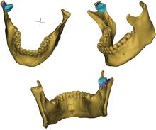
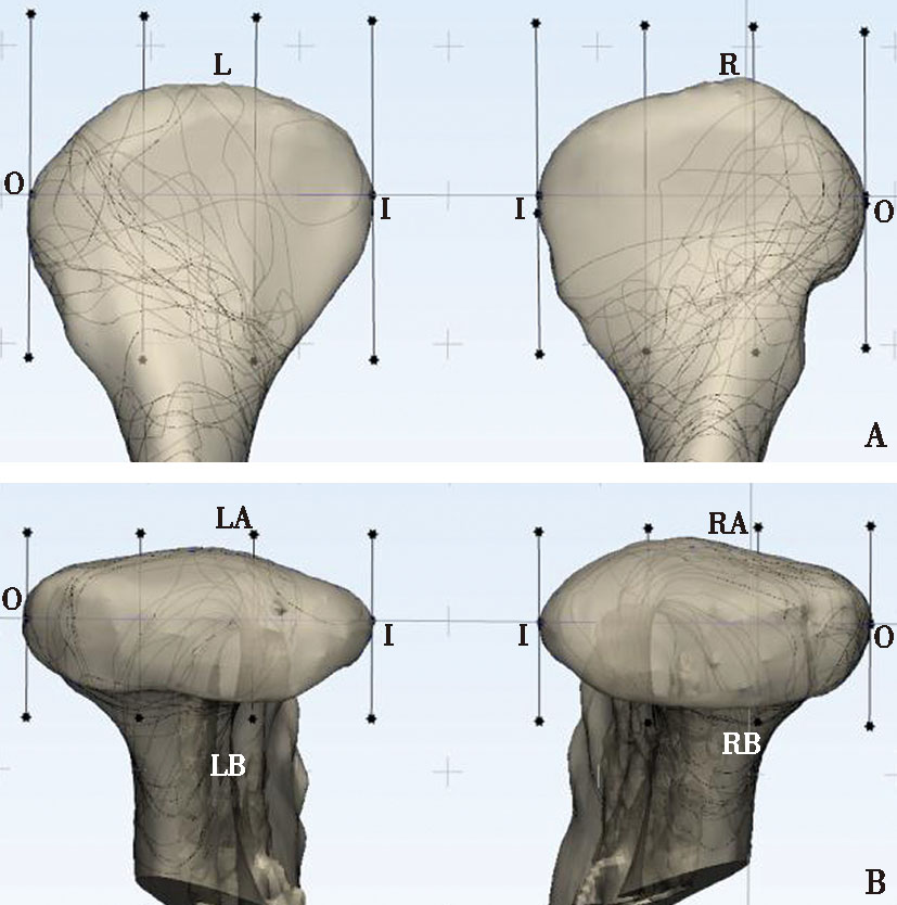
Fig.1
3D model of mandibular condyle in STL format"

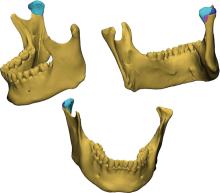
Fig.2
Resetting fracture breaks in 3-matic 16.0 software"
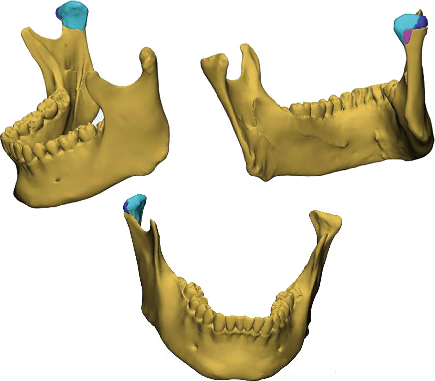
Fig.3
Adjusting transparency to copy the fracture line on the standard model"
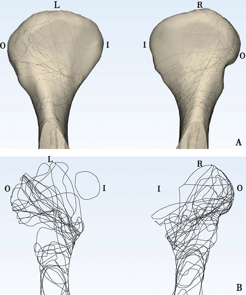
Fig.4
Delineation of condylar region"
Tab.1
Age distribution of spiral CT imaging data in 37 patients"
| 性别 | 18~20岁 | 21~30岁 | 31~40岁 | 41~50岁 | 51~60岁 | 60岁以上 |
|---|---|---|---|---|---|---|
| 女 | 0 | 5 | 4 | 1 | 0 | 1 |
| 男 | 1 | 6 | 7 | 6 | 6 | 0 |
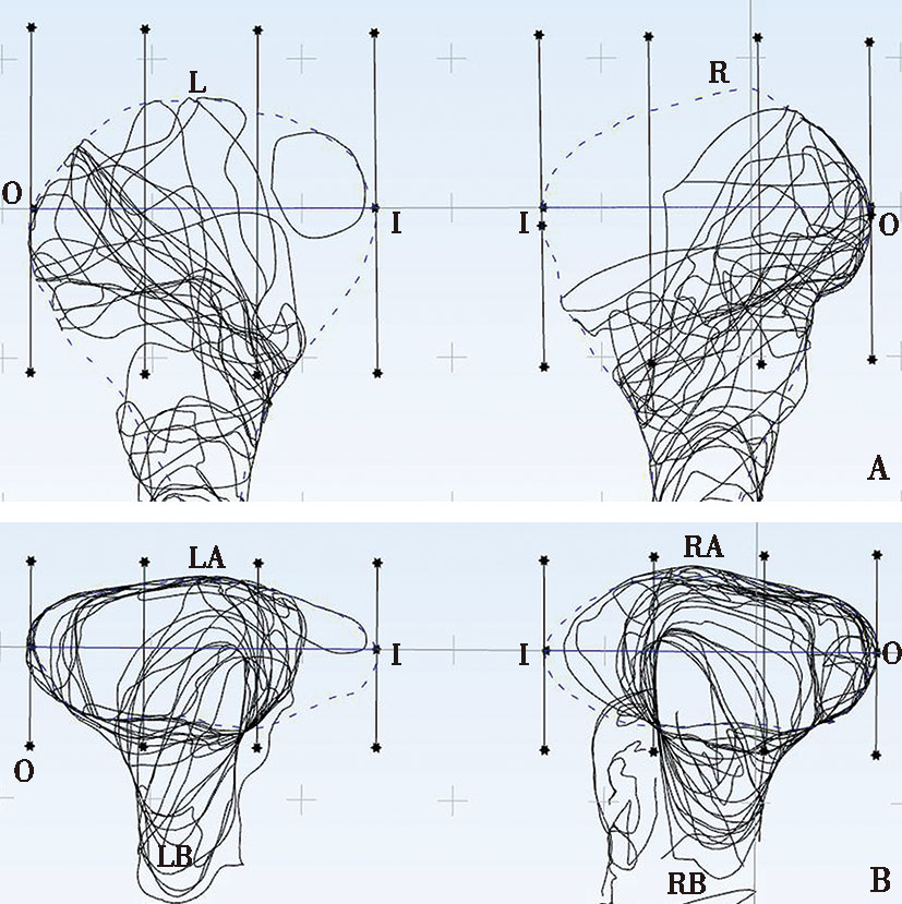
Fig.5
Fracture line alignment in 37 patients after hiding the standard mandible"
Tab.2
Distribution of condylar fracture lines in coronal position in 37 cases n(%)"
| 分部 | 左侧髁突骨折线分布 | 右侧髁突骨折线分布 | ||||
|---|---|---|---|---|---|---|
| 外部 | 中部 | 内部 | 外部 | 中部 | 内部 | |
| 上部 | 13(35.13) | 5(13.51) | 0 | 9(24.32) | 9(24.32) | 1(2.70) |
| 下部 | 16(43.24) | 17(45.94) | 14(37.83) | 7(18.92) | 12(32.43) | 12(32.43) |
Tab.3
Distribution of fracture lines in 37 sagittal condylar fractures n(%)"
| 分部 | 左侧髁突骨折线分布 | 右侧髁突骨折线分布 | ||||
|---|---|---|---|---|---|---|
| 外部 | 中部 | 内部 | 外部 | 中部 | 内部 | |
| 上部 | 17(45.94) | 12(32.43) | 7(18.92) | 13(35.13) | 10(27.03) | 3(8.10) |
| 下部 | 18(48.64) | 18(48.64) | 12(32.43) | 10(27.03) | 13(35.13) | 9(24.32) |

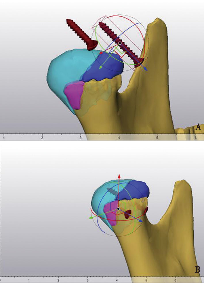
Fig.6
Imported threaded nail model"
| [1] |
Mooney S, Gulati RD, Yusupov S, et al. Mandibular condylar fractures[J]. Facial Plast Surg Clin North Am, 2022, 30(1):85-98.
doi: 10.1016/j.fsc.2021.08.007 |
| [2] | Nayak SS, Arun S, Taranath Kamath A, et al. The influence of the mandibular chin angle on the occurrence of mandibular condylar fracture: A retrospective study[J]. Sci World J, 2021, 2021:2380840. |
| [3] |
Shakya S, Zhang X, Liu L. Key points in surgical management of mandibular condylar fractures[J]. Chin J Traumatol, 2020, 23(2):63-70.
doi: S1008-1275(19)30181-6 pmid: 31744656 |
| [4] |
Bielecki-Kowalski B, Kozakiewicz M. Clinico-anatomical classification of the processus condylaris mandibulae for traumatological purposes[J]. Ann Anat, 2021, 234:151616.
doi: 10.1016/j.aanat.2020.151616 |
| [5] |
Bu LT, Chen Q, Huang K, et al. Evaluation of internal fixation techniques for condylar head fractures: A finite element analysis and comparison[J]. Oral Surg Oral Med Oral Pathol Oral Radiol, 2022, 133(5):e96-e104.
doi: 10.1016/j.oooo.2021.08.028 |
| [6] |
Arya R, Sritharan R, Glover S, et al. Non-surgical management of non-condylar mandibular fractures[J]. Br J Oral Maxillofac Surg, 2022, 60(9):1224-1227.
doi: 10.1016/j.bjoms.2022.07.014 |
| [7] |
Menon S, Kumar V, Archana S, et al. A retrospective study of condylar fracture management in a tertiary care hospital-a 10-year experience[J]. J Maxillofac Oral Surg, 2020, 19(3):380-386.
doi: 10.1007/s12663-019-01257-2 |
| [8] |
Nie W, Gu F, Wang ZJ, et al. Preliminary application of three-dimension printing technology in surgical management of bicondylar tibial plateau fractures[J]. Injury, 2019, 50(2):476-483.
doi: S0020-1383(18)30753-8 pmid: 30580928 |
| [9] |
King C, Shafi A, Burke E. Optimising the management of concurrent symphyseal/parasymphyseal and bilateral extracapsular condylar fractures using three-dimensional printing[J]. Oral Maxillofac Surg, 2020, 24(2):217-219.
doi: 10.1007/s10006-019-00820-y pmid: 31814066 |
| [10] | Kang HJ, Kim BS, Kim SM, et al. Can preoperative 3D printing change surgeon’s operative plan for distal Tibia fracture?[J]. Bio Med Res Int, 2019:1-7. |
| [11] |
Su QH, Zhang Y, Liao SH, et al. 3D computed tomography mapping of thoracolumbar vertebrae fractures[J]. Med Sci Monit, 2019, 25:2802-2810.
doi: 10.12659/MSM.915916 |
| [12] |
Xie XT, Zhan Y, Dong MJ, et al. Two and three-dimensional CT mapping of Hoffa fractures[J]. J Bone Joint Surg Am, 2017, 99(21):1866-1874.
doi: 10.2106/JBJS.17.00473 pmid: 29088042 |
| [13] |
He DM, Yang C, Chen MJ, et al. Intracapsular condylar fracture of the mandible: Our classification and open treatment experience[J]. J Oral Maxillofac Surg, 2009, 67(8):1672-1679.
doi: 10.1016/j.joms.2009.02.012 |
| [14] |
Thirunavukarasu AJ, Ferro A, Dubb SS, et al. Investigating the correlation between bone density and fracture frequency in the mandibular condyle with micro-computed tomography[J]. Br J Oral Maxillofac Surg, 2021, 59(3):380-383.
doi: 10.1016/j.bjoms.2020.08.097 pmid: 33495045 |
| [15] |
Jeyaraj P. Splitting and splaying apart of the craniomaxillofacial skeleton by medially directed disruptive forces of unusual etiologies-A case series[J]. Ann Maxillofac Surg, 2021, 11(1):195-213.
doi: 10.4103/ams.ams_275_20 pmid: 34522684 |
| [16] |
Yang RC, Cui MJ, Zhou HH, et al. Fracture fragment of the condyle determines the ramus height of the mandible in children with intracapsular condylar fractures treated conservatively[J]. Sci Rep, 2022, 12(1):19924.
doi: 10.1038/s41598-022-24463-4 |
| [17] | Tian SY, Liang SB, Wang ZZ, et al. Morphological characteristics of the posterior wall associated with complex acetabular fractures: A radiological study using 3D software and fracture mapping technique[J]. Bio Med Res Int, 2022:1-8. |
| [18] |
DeKeyser GJ, Sripanich Y, O’Neill DC, et al. Mapping of posterior talar dome access through posteromedial versus posterola-teral approaches[J]. J Orthop Trauma, 2021, 35(12):e463-e469.
doi: 10.1097/BOT.0000000000002113 pmid: 33724965 |
| [19] |
Zhou W, Rong QG, An JG, et al. Finite element analysis of two- and three-dimensional fixation in treating mandibular symphyseal fracture combined with bilateral condylar intracapsular fractures[J]. J Craniofac Surg, 2021, 32(7):2557-2561.
doi: 10.1097/SCS.0000000000007601 pmid: 33710062 |
| [20] |
Zhang C, Li ZB, Yang RT. Digital design and application of 3D printed surgical guide for long screw fixation of condylar sagittal fracture[J]. J Craniofac Surg, 2021, 32(7):e632-e634.
doi: 10.1097/SCS.0000000000007605 pmid: 33674507 |
| [21] |
Liokatis P, Tzortzinis G, Gerasimidis S, et al. Application of the lambda plate on condylar fractures: Finite element evaluation of the fixation rigidity for different fracture patterns and plate placements[J]. Injury, 2022, 53(4):1345-1352.
doi: 10.1016/j.injury.2022.01.032 pmid: 35101256 |
| [22] | Ben Achour A, Meißner H, Teicher U, et al. Biomechanical evaluation of mandibular condyle fracture osteosynthesis using the rhombic three-dimensional condylar fracture plate[J]. J Oral Maxillofac Surg, 2019, 77(9):1868.e1-1868.e15. |
| [23] |
Wang WH, Deng JY, Zhu J, et al. Computer-assisted virtual technology in intracapsular condylar fracture with two resorbable long-screws[J]. Br J Oral Maxillofac Surg, 2013, 51(2):138-143.
doi: 10.1016/j.bjoms.2012.04.005 pmid: 22546281 |
| [24] | 应凯, 王亮, 朱永武, 等. 虚拟手术结合导板辅助7例髁突颈部骨折复位固定的效果评价[J]. 上海口腔医学, 2023, 32(1):105-108. |
| [25] |
Johner JP, Essig H, Neff A, et al. Volumetric evaluated bone resorption after open reduction and internal fixation of condylar head fractures of the mandible[J]. J Oral Maxillofac Surg, 2021, 79(9):1902-1913.
doi: 10.1016/j.joms.2021.04.018 |
| [26] | 李豪培, 李金超, 李守宏. 下颌骨髁状突囊内骨折分类及手术治疗新进展[J]. 临床口腔医学杂志, 2020, 36(6):379-382. |
| Viewed | ||||||
|
Full text |
|
|||||
|
Abstract |
|
|||||