
口腔医学 ›› 2025, Vol. 45 ›› Issue (6): 436-439.doi: 10.13591/j.cnki.kqyx.2025.06.007
白煜1, 高盟2, 刘冬梅2, 王涛1, 冯雪3( )
)
收稿日期:2024-04-28
出版日期:2025-06-28
发布日期:2025-07-08
通讯作者:
冯雪 E-mail: 1054030184@qq.com
BAI Yu1, GAO Meng2, LIU Dongmei2, WANG Tao1, FENG Xue3()
Received:2024-04-28
Online:2025-06-28
Published:2025-07-08
摘要:
目的 通过三维数字化模型,对骨性Ⅱ类患者的下颌基骨弓与牙弓之间的关系进行研究,并与骨性Ⅰ类患者相对比,以期为临床准确诊断和后续治疗提供一定参考。方法 选取骨性Ⅱ类错牙合畸形患者的模型25副,骨性Ⅰ类个别正常牙合患者的模型25副,扫描选取的下颌模型并通过软件构建数字化三维模型。确定膜龈联合处最凸点并连线(WALA嵴),确定临床冠中心点(FA点),建立研究所需参考平面和平面直角坐标系。用WALA嵴点和FA点坐标进行四次多项式曲线拟合,代表相应的基骨弓曲线和牙弓曲线。在距离坐标水平轴3、10、18 mm处依次测量并计算基骨弓曲线与牙弓曲线的宽度差。比较骨性Ⅱ类与骨性Ⅰ类错牙合基骨弓和牙弓宽度差的差异。结果 骨性Ⅱ类患者下颌前段、中段及后段基骨弓与牙弓的宽度差分别为-1.58 mm、1.80 mm、3.80 mm,骨性Ⅰ类患者下颌前段、中段及后段基骨弓与牙弓的宽度差分别为2.08 mm、2.92 mm、4.24 mm,前段与中段骨性Ⅱ类与骨性Ⅰ相比较具差异有统计学意义(P<0.05),后段无统计学差异(P>0.05)。结论 骨性Ⅱ类患者前段牙弓位于基骨弓外侧,基骨弓宽度小于牙弓宽度;中段和后段牙弓位于基骨弓内侧,基骨弓宽度大于牙弓宽度。骨性Ⅰ类患者各段基骨弓宽度大于牙弓宽度,牙弓位于基骨内侧。骨性Ⅱ类患者中段基骨弓与牙弓的宽度差小于骨性Ⅰ类。
中图分类号:
白煜, 高盟, 刘冬梅, 王涛, 冯雪. 骨性Ⅱ类错牙合下颌基骨与牙弓形态的研究[J]. 口腔医学, 2025, 45(6): 436-439.
BAI Yu, GAO Meng, LIU Dongmei, WANG Tao, FENG Xue. Study on the morphology of the mandibular basal bone and dental arch of skeletal Class Ⅱ malocclusion[J]. Stomatology, 2025, 45(6): 436-439.
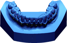
图1
确定参考点"

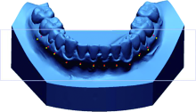
图2
建立参考平面"

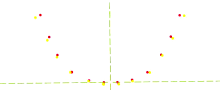
图3
建立平面直角坐标系"
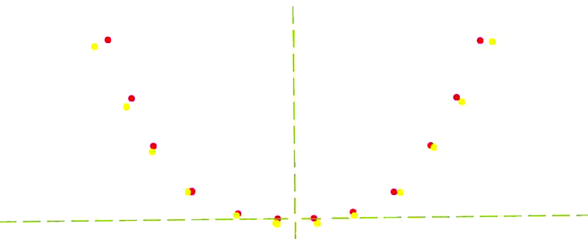
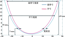
图4
宽度及宽度差测量"
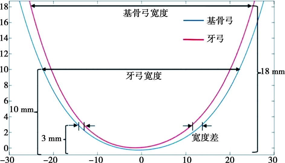
表1
骨性Ⅱ、Ⅰ类牙弓与基骨宽度"
| 分段 | 测量部位 | Ⅱ类 | Ⅰ类 | P |
|---|---|---|---|---|
| 前段 | 牙弓 | 27.12±2.53 | 23.54±2.49 | 0.000** |
| 基骨 | 25.54±1.73 | 25.61±1.34 | 0.887 | |
| 中段 | 牙弓 | 39.06±2.09 | 38.57±1.96 | 0.461 |
| 基骨 | 40.86±1.96 | 41.49±1.85 | 0.315 | |
| 后段 | 牙弓 | 46.86±2.73 | 47.70±2.63 | 0.343 |
| 基骨 | 50.67±2.22 | 51.94±2.43 | 0.095 |
表2
骨性Ⅱ类、Ⅰ类下颌基骨与牙弓的宽度差"
| 分段 | 宽度差 | t | P | |
|---|---|---|---|---|
| 前段 | Ⅱ类 | -1.58±2.44 | 4.527 | 0.000* |
| Ⅰ类 | 2.08±2.59 | |||
| 中段 | Ⅱ类 | 1.80±1.49 | 2.298 | 0.027* |
| Ⅰ类 | 2.92±1.54 | |||
| 后段 | Ⅱ类 | 3.80±1.42 | 1.011 | 0.318 |
| Ⅰ类 | 4.24±1.20 |
| [1] | 傅民魁, 张丁, 王邦康, 等. 中国25 392名儿童与青少年错牙合畸形患病率的调查[J]. 中华口腔医学杂志, 2002, 37(5): 371-373. |
| [2] | Ronay V, Miner RM, Will LA, et al. Mandibular arch form: The relationship between dental and basal anatomy[J]. Am J OrthodDentofacial Orthop, 2008, 134(3): 430-438. |
| [3] | Kim KY, Bayome M, Kim K, et al. Three-dimensional evaluation of the relationship between dental and basal arch forms in normal occlusion[J]. Korean J Orthod, 2011, 41(4): 288. |
| [4] | 奚祺, 吴国锋. 数字化口内扫描技术的发展与应用[J]. 实用口腔医学杂志, 2021, 37(1): 136-140. |
| [5] | Hayama K, Arai K, Ishikawa H. Correlation between upper and lower dental arch froms by fitting of fourth-order polynomials[J]. Orthod Waves, 2000, 59:303-311. |
| [6] | 高盟. 骨性Ⅱ类错牙合拔牙治疗前后基骨与牙弓形态的研究[D]. 西安: 中国人民解放军空军军医大学, 2018. |
| [7] |
Wen YF, Wong HM, Pei T, et al. Adolescent dental arch development among southern Chinese in Hong Kong: A geometric morphometric approach[J]. Sci Rep, 2019, 9(1): 18526.
doi: 10.1038/s41598-019-55073-2 pmid: 31811230 |
| [8] | 高盟, 刘冬梅, 程锦, 等. 骨性Ⅱ类错牙合拔牙矫治前后下颌牙弓与基骨形态的变化[J]. 中华医学美学美容杂志, 2018, 24(3): 188-191. |
| [9] | Brash JC. The etiology of irregularity and malocclusion of the teeth[M]. London: Dental Board of the United Kingdom, 1956. |
| [10] | Andrews L. The six elements of orofacial harmony[J]. Andrews J, 2000(1):13-22. |
| [11] | Ramón R, Adanero A, Miegimolle M. A new approach to diagnosis to posterior cross bite: Intraoral photography and walaridge[J]. Int J Environ Res Public Health, 2022, 19(15): 9443. |
| [12] | Uysal T, Badel M, Serdar U, et al. Dental and alveolar arch widths in normal occlusion, Class Ⅱ division 1 and Class Ⅱ division 2[J]. Angle Orthod, 2005, 75(6): 941-947. |
| [13] | Gupta D, Miner RM, Arai K, et al. Comparison of the mandibular dental and basal arch forms in adults and children with Class Ⅰ and Class Ⅱmalocclusions[J]. Am J Orthod Dentofacial Orthop, 2010, 138(1): 10. e1-10. e8;discussion 10-11. |
| [14] |
Ueno K, Kumabe S, Nakatsuka M, et al. Factors influencing dental arch form[J]. Okajimas Folia Anat Jpn, 2019, 96(1): 31-46.
doi: 10.2535/ofaj.96.31 pmid: 31462623 |
| [15] | 李蓉, 戴宁, 陈嵘, 等. 安氏Ⅱ1类错牙合与正常牙合牙弓宽度的研究[J]. 临床口腔医学杂志, 2009, 25(11): 677-679. |
| [16] | Buschang PH, Roldan SI, Tadlock LP. Guidelines for assessing the growth and development of orthodontic patients[J]. Semin Orthod, 2017, 23(4): 321-335. |
| [17] | 徐舒豪, 彭薇, 黄诗言, 等. 不同矢状骨性错牙合畸形牙弓及基骨宽度的比较研究[J]. 成都医学院学报, 2024, 19(1): 28-33. |
| [18] | 陈上, 厉松. 自锁托槽解除上颌牙列拥挤后牙弓与牙齿的三维变化研究[J]. 中华口腔正畸学杂志, 2016, 23(2): 67-72. |
| [1] | 陈子昂, 刘子昂, 欧阳雨晴, 娄依婷, 施洁珺, 丁王辉. 正畸治疗中周围解剖结构与牙根外吸收之间关系的综述[J]. 口腔医学, 2025, 45(5): 394-400. |
| [2] | 闫行之, 蔡新誉, 陈思麦, 方韦文, 雷凡, 曹丹, 张阳. PAD4在颌骨生长发育调控中的作用初探[J]. 口腔医学, 2025, 45(4): 241-247. |
| [3] | 房斌, 李媛, 周薇娜, 于林凤, 周猛, 马俊青. 无托槽隐形矫治初期温度感觉与压力痛觉的研究[J]. 口腔医学, 2025, 45(4): 259-263. |
| [4] | 黎金鹏, 江银华. 儿童腺样体肥大与错𬌗畸形相关性的研究进展[J]. 口腔医学, 2025, 45(4): 311-316. |
| [5] | 苏咏宽, 潘永初, 张晶超, 卞海峰, 方玉心, 侯伟, 韩霖霏. 上颌前方牵引对替牙期唇腭裂患者软硬组织的影响[J]. 口腔医学, 2025, 45(3): 168-174. |
| [6] | 赵玺, 杨莉. 隐形矫治下颌尖牙移动最佳附件位置的三维有限元分析[J]. 口腔医学, 2025, 45(3): 197-203. |
| [7] | 刘珂, 轩诗杰, 刘鑫. 颧牙槽嵴支抗稳定性影响因素的三维有限元分析[J]. 口腔医学, 2025, 45(2): 100-104. |
| [8] | 马惠, 王雯, 仇岩, 华泽权. 下颌支矢状骨劈开术后退下颌治疗骨性Ⅲ类错𬌗畸形对上气道和睡眠呼吸通气功能的影响[J]. 口腔医学, 2025, 45(2): 123-128. |
| [9] | 包佳琦, 王中秀, 冯贻苗, 雷利红, 陈莉丽. 正畸治疗中牙周硬组织相关并发症的处理[J]. 口腔医学, 2025, 45(1): 37-44. |
| [10] | 李根, 王华, 谷妍. 数字化三维打印前方牵引联合快速扩缩矫治替牙期骨性Ⅲ类的临床效果研究[J]. 口腔医学, 2025, 45(1): 51-57. |
| [11] | 周悦, 唐振兴, 李宇. 拔除前磨牙正畸病例的磨牙支抗丧失及其影响因素[J]. 口腔医学, 2025, 45(1): 64-68. |
| [12] | 程烨, 曾维浩, 李煌, 雷浪. 下颌第二磨牙远中移动牙根限制相关因素的锥形束计算机断层扫描研究[J]. 口腔医学, 2024, 44(12): 887-891. |
| [13] | 王若飞, 邵丽鑫, 刘晓彤, 张苗苗. GsMTx4对大鼠牙齿移动过程中痛觉的抑制作用[J]. 口腔医学, 2024, 44(11): 815-819. |
| [14] | 李宗峰, 孙莲, 潘永初. 不同矢状骨面型成人下颌磨牙远中舌侧间隙的CBCT研究[J]. 口腔医学, 2024, 44(11): 820-823. |
| [15] | 娄姝, 采晓燕, 张驰, 张元, 韩旻轩, 管兆兰. 比较无托槽隐形和固定矫治对青少年患者生活质量的影响[J]. 口腔医学, 2024, 44(11): 841-846. |
| 阅读次数 | ||||||
|
全文 |
|
|||||
|
摘要 |
|
|||||
 苏公网安备32010602011670号