
Stomatology ›› 2024, Vol. 44 ›› Issue (8): 590-595.doi: 10.13591/j.cnki.kqyx.2024.08.007
• Basic and Clinical Research • Previous Articles Next Articles
ZHENG Yao1,2,YU Shaoyang1,2,HUANG Wenli1,2,DONG Lirong1,2,ZHANG Qiang1,YUAN Xiao1,2( )
)
Received:2024-01-11
Online:2024-08-28
Published:2024-08-06
CLC Number:
ZHENG Yao, YU Shaoyang, HUANG Wenli, DONG Lirong, ZHANG Qiang, YUAN Xiao. Three-dimensional evaluation of changes in the upper airway, natural head position and hyoid bone position in skeletal Class Ⅲ patients after bimaxillary surgery[J]. Stomatology, 2024, 44(8): 590-595.
Tab.1
Landmarks"
| 参考点 | 定义 |
|---|---|
| Na | 鼻额缝的最前点 |
| Ba | 枕骨大孔前缘中点 |
| Se | 蝶鞍中心点 |
| OrR | 右侧眶下缘中点 |
| PoL | 左侧外耳道最上点 |
| PoR | 右侧外耳道最上点 |
| Roof | 鼻咽顶部 |
| PNS | 硬腭的最后点 |
| A | 前鼻棘与上牙槽缘点之间的骨部最凹点 |
| B | 下牙槽突缘点与颏前点之间的骨部最凹点 |
| H | 舌骨最上最前点 |
| C2s | 第二颈椎上后极切点 |
| C2i | 第二颈椎下后点 |
| C4i | 第四颈椎下后点 |
| CV2 | 第二颈椎下前点 |
| CV3 | 第三颈椎下前点 |
| CV4 | 第四颈椎下前点 |
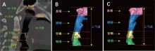
Fig.1
3D reconstruction of upper airway segments"

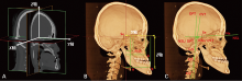
Fig.2
3D measurement schematic"

Tab.2
Surgical changes in the skeletal position and cranio-cervical angle"
| 变量 | T0 | T1 | T2 | T1-T0 | P | T2-T0 | P | T2-T1 | P | |
|---|---|---|---|---|---|---|---|---|---|---|
| 骨骼位置 | ||||||||||
| PNS/mm | Y轴 | 18.20±2.67 | 20.58±3.42 | 20.26±3.41 | 2.38±2.30 | 0.003** | 2.06±2.21 | 0.003** | -0.32±0.38 | 0.064 |
| Z轴 | 44.05±3.34 | 44.15±3.36 | 44.22±3.15 | 0.10±1.05 | 0.694 | 0.17±0.70 | 0.272 | 0.07±0.55 | 0.813 | |
| A/mm | Y轴 | 63.51±3.62 | 65.43±3.87 | 65.16±3.96 | 1.92±2.16 | 0.015* | 1.66±2.04 | 0.041* | -0.26±0.46 | 0.075 |
| Z轴 | 51.89±5.62 | 51.77±5.31 | 51.74±5.40 | -0.13±2.54 | 0.875 | -0.14±2.34 | 0.753 | -0.02±0.72 | 0.637 | |
| B/mm | Y轴 | 69.06±5.75 | 64.04±6.58 | 64.35±6.74 | -5.03±4.25 | 0.005** | -4.71±4.05 | 0.005** | 0.31±0.95 | 0.308 |
| Z轴 | 95.25±8.43 | 93.83±6.84 | 93.56±7.26 | -1.41±4.22 | 0.239 | -1.68±3.01 | 0.099 | -0.27±1.50 | 0.480 | |
| H/mm | Y轴 | 19.35±6.80 | 14.95±6.27 | 15.85±6.32 | -4.39±3.80 | 0.006** | -3.50±2.51 | 0.005** | 0.90±1.50 | 0.099 |
| Z轴 | 98.44±10.14 | 105.15±11.08 | 103.89±11.34 | 6.71±4.00 | 0.003** | 5.46±3.93 | 0.003** | -1.26±0.73 | 0.003** | |
| 颅颈角 | ||||||||||
| NSL/OPT/(°) | 99.06±6.02 | 101.44±6.15 | 102.80±5.59 | 2.38±2.73 | 0.015* | 3.64±2.67 | 0.005** | 1.25±1.80 | 0.031* | |
| NSL/CVT/(°) | 101.92±4.43 | 104.35±4.83 | 104.70±4.72 | 2.43±2.06 | 0.005 | 2.79±1.91 | 0.003 | 0.36±1.74 | 0.197 |
Tab.3
Surgical changes in upper airway volume"
| 变量 | T0 | T1 | T2 | T1-T0 | P | T2-T0 | P | T2-T1 | P |
|---|---|---|---|---|---|---|---|---|---|
| NP/mm3 | 8 226.89± 2 707.39 | 7 710.40± 3 356.70 | 8 124.25± 3 051.18 | -611.80± 1415.68 | 0.182 | -197.94± 879.80 | 0.328 | 413.85± 695.61 | 0.062 |
| PP/mm3 | 14 598.61± 5 386.37 | 11 217.05± 4 080.98 | 11 808.52± 4 032.12 | -3 381.55± 2 807.76 | 0.003** | -2 790.09± 2 358.70 | 0.003** | 591.46± 664.30 | 0.005** |
| GP/mm3 | 6 417.16± 2 036.86 | 5 335.74± 1 162.28 | 5 571.05± 1 284.02 | -1 081.41± 1 294.44 | 0.008** | -846.11± 1029.61 | 0.008** | 235.30± 295.57 | 0.023* |
| HP/mm3 | 6 262.70± 2 138.28 | 5 497.59± 1 600.29 | 5 697.34± 1 497.32 | -765.10± 1913.80 | 0.239 | -565.36± 1446.35 | 0.347 | 199.74± 514.00 | 0.209 |
| TP/mm3 | 35 697.66± 11 382.63 | 29 550.84± 8 905.01 | 31 053.32± 9 013.40 | -6 146.82± 5 991.69 | 0.002** | -4 644.33± 4 960.28 | 0.003** | 1 502.48± 1 209.72 | 0.002** |
Tab.4
Correlation of changes in maxilla-mandible position and cranio-cervical angle with changes in upper airway volume and hyoid position"
| 变量 | ΔNP | ΔPP | ΔGP | ΔHP | ΔTP | H-ΔY | H-ΔZ | ||||||||||||||
|---|---|---|---|---|---|---|---|---|---|---|---|---|---|---|---|---|---|---|---|---|---|
| T1-T0 | T2-T0 | T1-T0 | T2-T0 | T1-T0 | T2-T0 | T1-T0 | T2-T0 | T1-T0 | T2-T0 | T1-T0 | T2-T0 | T1-T0 | T2-T0 | ||||||||
| 上下颌骨位置变化 | |||||||||||||||||||||
| PNS-ΔY | -0.015 | 0.109 | -0.019 | 0.110 | 0.152 | 0.260 | 0.118 | 0.218 | 0.087 | 0.251 | 0.165 | 0.136 | -0.169 | 0.137 | |||||||
| PNS-ΔZ | 0.338 | 0.109 | 0.025 | 0.007 | -0.141 | -0.044 | 0.004 | 0.083 | 0.031 | 0.056 | 0.012 | -0.018 | 0.222 | -0.019 | |||||||
| A-ΔY | -0.033 | -0.047 | 0.490 | 0.453 | 0.529 | 0.440 | 0.480 | 0.436 | 0.552 | 0.577 | -0.401 | -0.553 | -0.421 | -0.553 | |||||||
| A-ΔZ | -0.262 | -0.242 | 0.542 | 0.311 | 0.451 | 0.231 | 0.158 | 0.060 | 0.325 | 0.051 | -0.048 | 0.149 | 0.222 | 0.149 | |||||||
| B-ΔY | -0.150 | -0.189 | 0.402 | 0.434 | 0.462 | 0.509 | 0.466 | 0.497 | 0.586* | 0.621* | -0.496 | -0.455 | -0.577 | -0.455 | |||||||
| B-ΔZ | -0.243 | -0.299 | 0.546 | 0.507 | 0.421 | 0.447 | 0.118 | 0.218 | 0.169 | 0.189 | 0.092 | 0.089 | 0.029 | 0.089 | |||||||
| 颅颈角变化 | |||||||||||||||||||||
| NSL/OPT | 0.046 | 0.271 | 0.430 | 0.544 | 0.054 | 0.423 | -0.226 | 0.323 | 0.176 | 0.468 | -0.084 | 0.002 | 0.337 | 0.002 | |||||||
| NSL/CVT | -0.565 | -0.097 | 0.300 | 0.594* | 0.172 | 0.615* | -0.000 | 0.547 | 0.001 | 0.431 | 0.037 | -0.253 | 0.183 | -0.254 | |||||||
| [1] | Kang NE, Lee DH, Seo JI, et al. Postoperative changes in the pharyngeal airway space through computed tomography evaluation after mandibular setback surgery in skeletal Class Ⅲ patients: 1-year follow-up[J]. Maxillofac Plast Reconstr Surg, 2021, 43(1): 31. |
| [2] |
郭蕾, 康非吾, 陈袁伟. 正颌手术患者报告结局量表的研究现状[J]. 口腔颌面外科杂志, 2022, 32(5): 315-318.
doi: 10.3969/j.issn.1005-4979.2022.05.009 |
| [3] | Tseng YC, Hsiao SY, Cheng JH, et al. Postoperative skeletal stability and pharyngeal airway: Counterclockwise versus clockwise rotation during mandibular setback surgery[J]. Biomed Res Int, 2020: 3283080. |
| [4] | Kim SH, Choi SK. Changes in the hyoid bone, tongue, and oropharyngeal airway space after mandibular setback surgery evaluated by cone-beam computed tomography[J]. Maxillofac Plast Reconstr Surg, 2020, 42(1): 27. |
| [5] | On SW, Kim HJ, Cho DH, et al. Silent changes in sleep quality following mandibular setback surgery in patients with skeletal Class Ⅲ malocclusion: A prospective study[J]. Sci Rep, 2019, 9(1): 9737. |
| [6] | Yang HJ, Jung YE, Kwon IJ, et al. Airway changes and prevalence of obstructive sleep apnoea after bimaxillary orthognathic surgery with large mandibular setback[J]. Int J Oral Maxillofac Surg, 2020, 49(3): 342-349. |
| [7] | Kim MA, Kim BR, Youn JK, et al. Head posture and pharyngeal airway volume changes after bimaxillary surgery for mandibular prognathism[J]. J Craniomaxillofac Surg, 2014, 42(5): 531-535. |
| [8] |
Lin XZ. Correlation study of increase of pharyngeal airway space after mandibular advancement, taking natural head position into consideration[J]. Br J Oral Maxillofac Surg, 2019, 57(8): 760-764.
doi: S0266-4356(19)30268-2 pmid: 31345578 |
| [9] | 余紫嘉, 郑之峻. 自然头位在正畸领域的应用[J]. 实用临床医药杂志, 2021, 25(12): 113-118. |
| [10] | Achilleos S, Krogstad O, Lyberg T. Surgical mandibular setback and changes in uvuloglossopharyngeal morphology and head posture: A short- and long-term cephalometric study in males[J]. Eur J Orthod, 2000, 22(4): 383-394. |
| [11] |
Zheng LY, Jahn J, Vasavada AN. Sagittal plane kinematics of the adult hyoid bone[J]. J Biomech, 2012, 45(3): 531-536.
doi: 10.1016/j.jbiomech.2011.11.040 pmid: 22176712 |
| [12] | Samaha CJ, Tannous HJ, Salman D, et al. Role of surgical hyoid bone repositioning in modifying upper airway collapsibility[J]. Front Physiol, 2022, 13: 1089606. |
| [13] |
Park JH, Kim HS, Choi SH, et al. Changes in position of the hyoid bone and volume of the pharyngeal airway after mandibular setback: Three-dimensional analysis[J]. Br J Oral Maxillofac Surg, 2019, 57(1): 29-35.
doi: S0266-4356(18)30674-0 pmid: 30598316 |
| [14] | Jiang YY. Correlation between hyoid bone position and airway dimensions in Chinese adolescents by cone beam computed tomography analysis[J]. Int J Oral Maxillofac Surg, 2016, 45(7): 914-921. |
| [15] | Jeong S, Sung J, Kim S, et al. Upper airway morphologic changes after mandibular setback surgery in skeletal Class Ⅲ malocclusion patients measured using cone beam computed tomography superimposition[J]. Int J Oral Maxillofac Surg, 2018, 47(11): 1405-1410. |
| [16] | Kang Y, Lee S, Gong Y, et al. Three-dimensional morphologic evaluation of the changes in the pharyngeal airway and hyoid bone after bimaxillary surgery in patients with skeletal Class Ⅲ malocclusion with facial asymmetry: A preliminary study[J]. Am J Orthod Dentofacial Orthop, 2022, 162(1): 42-50. |
| [17] | Tan SK, Tang ATH, Leung WK, et al. Three-dimensional pharyngeal airway changes after 2-jaw orthognathic surgery with segmentation in dento-skeletal Class Ⅲ patients[J]. J Craniofac Surg, 2019, 30(5): 1533-1538. |
| [18] | An JH, Park SB, Choi YK, et al. Cone-beam computed tomography evaluation of pharyngeal airway space changes after bimaxillary orthognathic surgery in patients with Class Ⅲ skeletal deformities: A 6-year follow-up study[J]. J Oral Maxillofac Surg, 2019, 77(12): 2534-2544. |
| [19] | Jakobsone G, Stenvik A, Espeland L. The effect of maxillary advancement and impaction on the upper airway after bimaxillary surgery to correct Class Ⅲ malocclusion[J]. Am J Orthod Dentofacial Orthop, 2011, 139(4 Suppl): e369-e376. |
| [20] | Giap HV, Shin JW, Chae HS, et al. Pharyngeal airway morphology in skeletal Class Ⅲ with mandibular asymmetry is improved after bimaxillary orthognathic surgery[J]. J Oral Maxillofac Surg, 2021, 79(5): 1107-1121. |
| [21] | Yang YX, Yang K, Zhao Y. Three-dimensional changes in the upper airway of skeletal Class Ⅲ patients after different orthognathic surgical procedures[J]. J Oral Maxillofac Surg, 2018, 76(1): 155-164. |
| [22] | Souza Pinto GN, Iwaki Filho L, Previdelli ITDS, et al. Three-dimensional alterations in pharyngeal airspace, soft palate, and hyoid bone of Class Ⅱ and Class Ⅲ patients submitted to bimaxillary orthognathic surgery: A retrospective study[J]. J Craniomaxillofac Surg, 2019, 47(6): 883-894. |
| [23] | Khaghaninejad MS, Khojastehpour L, Danesteh H, et al. Changes in the pharyngeal airway after different orthognathic procedures for correction of Class Ⅲ dysplasia[J]. Maxillofac Plast Reconstr Surg, 2022, 44(1): 23. |
| [24] |
Eggensperger N, Smolka W, Iizuka T. Long-term changes of hyoid bone position and pharyngeal airway size following mandibular setback by sagittal split ramus osteotomy[J]. J Craniomaxillofac Surg, 2005, 33(2): 111-117.
pmid: 15804590 |
| [25] |
Bibby RE, Preston CB. The hyoid triangle[J]. Am J Orthod, 1981, 80(1): 92-97.
doi: 10.1016/0002-9416(81)90199-8 pmid: 6942659 |
| Viewed | ||||||
|
Full text |
|
|||||
|
Abstract |
|
|||||