
Stomatology ›› 2023, Vol. 43 ›› Issue (6): 524-528.doi: 10.13591/j.cnki.kqyx.2023.06.008
• Clinical Research • Previous Articles Next Articles
ZHANG Chengcheng,ZHOU Xi,FAN Liwen,MA Lan,PAN Yongchu( )
)
Revised:2023-03-15
Online:2023-06-28
Published:2023-07-06
CLC Number:
ZHANG Chengcheng, ZHOU Xi, FAN Liwen, MA Lan, PAN Yongchu. A comparative study on morphological characteristics of condyle-fossa for skeletal Class Ⅰ and Ⅲ malocclusion[J]. Stomatology, 2023, 43(6): 524-528.
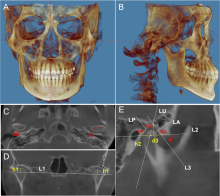
Fig.1
Correction of head position and related detection indicators of temporomandibular joint"

Tab.1
Difference analysis of measurements between the left and right TMJ among the three groups"
| 测量指标 | 骨性Ⅰ类 | 上颌骨发育不足 | 下颌骨发育过度 | |||||||
|---|---|---|---|---|---|---|---|---|---|---|
| 左侧 | 右侧 | P* | 左侧 | 右侧 | P* | 左侧 | 右侧 | P* | ||
| 髁突上部高度/mm | 5.12±1.11 | 5.01±0.87 | 0.463 | 6.32±1.87 | 6.52±1.64 | 0.556 | 6.32±1.13 | 6.60±1.63 | 0.235 | |
| 髁突内外嵴/mm | 18.02±1.74 | 18.45±2.25 | 0.308 | 18.07±2.24 | 18.21±2.39 | 0.640 | 18.14±1.85 | 18.52±2.01 | 0.214 | |
| 髁突前后嵴/mm | 7.55±0.84 | 7.54±0.81 | 0.965 | 7.05±1.17 | 7.29±1.15 | 0.247 | 7.51±1.03 | 7.39±1.03 | 0.618 | |
| 关节结节后斜面倾斜度/(°) | 36.86±5.27 | 35.68±6.17 | 0.454 | 32.96±6.82 | 34.97±11.87 | 0.350 | 31.75±7.92 | 30.79±7.04 | 0.561 | |
| 关节窝高度/mm | 7.40±0.97 | 7.42±0.94 | 0.934 | 7.01±1.01 | 7.08±1.11 | 0.704 | 6.79±1.61 | 6.74±1.34 | 0.839 | |
| 关节窝宽度/mm | 19.49±1.61 | 19.84±1.28 | 0.288 | 20.0±2.34 | 19.59±2.50 | 0.245 | 20.84±2.41 | 20.46±2.22 | 0.422 | |
Tab.2
Difference analysis of measurements between the left and right TMJ among the three groups"
| 测量指标 | 骨性Ⅰ类 | 上颌骨发育不足 | 下颌骨发育过度 | |||||||
|---|---|---|---|---|---|---|---|---|---|---|
| 左侧 | 右侧 | P* | 左侧 | 右侧 | P* | 左侧 | 右侧 | P* | ||
| 关节上间隙/mm | 4.98 (4.30~5.28) | 3.55 (3.39~4.09) | 0.478 | 4.02 (3.35~4.87) | 3.28 (2.66~3.82) | 0.177 | 3.91 (3.20~4.74) | 3.56 (2.74 ~ 4.14) | 0.911 | |
| 关节前间隙/mm | 3.76 (3.36~4.39) | 4.81 (4.51~5.15) | 0.067 | 3.76 (3.26~4.20) | 4.22 (3.59~5.57) | 0.240 | 3.73 (3.22~4.50) | 4.12 (3.31~ 4.49) | 0.573 | |
| 关节后间隙/mm | 3.26 (3.09~3.84) | 3.62 (3.16~4.09) | 0.232 | 3.32 (2.76~4.61) | 3.64 (3.03~4.05) | 0.297 | 3.88 (2.98 ~ 4.17) | 3.86 (3.42~ 4.02) | 0.940 | |
Tab.3
Difference analysis of measurements among the three groups"
| 测量指标 | 骨性Ⅰ类 | 上颌骨发育不足 | 下颌骨发育过度 | P1 | P2 | P3 |
|---|---|---|---|---|---|---|
| 髁突上部高度/mm | 5.07± 0.99 | 6.42±1.74 | 6.47±1.40 | 0.000* | 0.000* | 0.999 |
| 关节前间隙/mm | 3.63±0.66 | 3.58±1.22 | 3.61±0.86 | 0.790 | 0.870 | 0.920 |
| 关节后间隙/mm | 3.71±0.66 | 3.62±0.72 | 3.81±0.64 | 0.564 | 0.528 | 0.235 |
| 髁突内外嵴/mm | 18.24±2.00 | 18.14±2.38 | 18.33±1.92 | 0.842 | 0.840 | 0.692 |
| 髁突前后嵴/mm | 7.54±0.81 | 7.17±1.15 | 7.45±1.02 | 0.106 | 0.674 | 0.226 |
| 关节窝高度/mm | 7.41±0.95 | 7.04±1.05 | 6.76±1.46 | 0.175 | 0.016* | 0.307 |
Tab.4
Difference analysis of measurements among the three groups"
| 测量指标 | 骨性Ⅰ类 | 上颌骨发育不足 | 下颌骨发育过度 | P |
|---|---|---|---|---|
| 关节上间隙/mm | 3.47(3.19~4.01) | 3.32(2.73~4.12) | 3.645(2.92~4.14) | 0.505 |
| 关节结节后斜面倾斜度/(°) | 36.60(32.53~40.06) | 32.68(27.32~37.26) | 31.19(26.05~36.23) | 0.004** |
| 关节窝宽度/mm | 19.79(18.94~20.83) | 19.78(17.50~21.64) | 20.15(19.07~21.72) | 0.183 |
| [1] |
Al-Hadad SA, ALyafrusee ES, Abdulqader AA, et al. Comprehensive three-dimensional positional and morphological assessment of the temporomandibular joint in skeletal Class Ⅱ patients with mandibular retrognathism in different vertical skeletal patterns[J]. BMC Oral Health, 2022, 22(1):149.
doi: 10.1186/s12903-022-02174-6 pmid: 35484618 |
| [2] | 董春梅, 俞律峰, 邹德荣. 错𬌗畸形患者颞下颌关节的CBCT研究进展[J]. 口腔医学, 2020, 40(6):560-564. |
| [3] |
Arieta-Miranda JM, Silva-Valencia M, Flores-Mir C, et al. Spatial analysis of condyle position according to sagittal skeletal relationship, assessed by cone beam computed tomography[J]. Prog Orthod, 2013, 14: 36.
doi: 10.1186/2196-1042-14-36 pmid: 24325842 |
| [4] |
Schiffman E, Ohrbach R, Truelove E, et al. Diagnostic criteria for temporomandibular disorders(DC/TMD)for clinical and research applications: Recommendations of the international RDC/TMD consortium network and orofacial pain special interest group[J]. J Oral Facial Pain Headache, 2014, 28(1):6-27.
doi: 10.11607/jop.1151 |
| [5] | Lin M, Xu YF, Wu HR, et al. Comparative cone-beam computed tomography evaluation of temporomandibular joint position and morphology in female patients with skeletal Class Ⅱ malocclusion[J]. J Int Med Res, 2019, 48(2):300060519892388. |
| [6] | Zhou X, Zhang CC, Yao SY, et al. Genetic architecture of non-syndromic skeletal Class Ⅲ malocclusion[J]. Oral Dis, 2022:2022, 23(18):10673. |
| [7] |
Yao SY, Zhou X, Vona B, et al. Skeletal Class Ⅲ malocclusion is associated with ADAMTS2 variants and reduced expression in a familial case[J]. Int J Mol Sci, 2022, 23(18):10673.
doi: 10.3390/ijms231810673 |
| [8] |
Zhang ZL, Cheng JG, Li G, et al. Measurement accuracy of temporomandibular joint space in Promax 3-dimensional cone-beam computerized tomography images[J]. Oral Surg Oral Med Oral Pathol Oral Radiol, 2012, 114(1):112-117.
doi: 10.1016/j.oooo.2011.11.020 |
| [9] |
Hasebe A, Yamaguchi T, Nakawaki T, et al. Comparison of condylar size among different anteroposterior and vertical skeletal patterns using cone-beam computed tomography[J]. Angle Orthod, 2019, 89(2):306-311.
doi: 10.2319/032518-229.1 pmid: 30475648 |
| [10] | Nithin 1 Ahmed J, Sujir N, et al. Morphological assessment of TMJ spaces, mandibular condyle, and glenoid Fossa using cone beam computed tomography(CBCT):A retrospective analysis[J]. Indian J Radiol Imaging, 2021, 31(1):78-85. |
| [11] | 王丹, 方梦如, 李强, 等. 正畸正颌联合治疗骨性Ⅲ类错𬌗畸形颞下颌关节变化[J]. 口腔生物医学, 2020, 11(3):176-180. |
| [12] | 牟婷琛, 冯剑颖, 章振兴. Mimics在颞下颌关节紊乱病髁突体积、表面积及形态学指数测量中的应用[J]. 口腔医学, 2022, 42(12):1097-1100. |
| [13] | 武杰. 成人高角骨性Ⅲ类错𬌗和成人正常𬌗颞下颌关节的CBCT对比研究[D]. 天津: 天津医科大学, 2012. |
| [14] |
Liu Q, Wei XE, Guan JJ, et al. Assessment of condylar morphology and position using MSCT in an Asian population[J]. Clin Oral Investig, 2018, 22(7):2653-2661.
doi: 10.1007/s00784-018-2364-7 |
| [15] |
Chae JM, Park JH, Tai K, et al. Evaluation of condyle-fossa relationships in adolescents with various skeletal patterns using cone-beam computed tomography[J]. Angle Orthod, 2020, 90(2):224-232.
doi: 10.2319/052919-369.1 |
| [16] |
Burke G, Major P, Glover K, et al. Correlations between condylar characteristics and facial morphology in Class Ⅱ preadolescent patients[J]. Am J Orthod Dentofacial Orthop, 1998, 114(3):328-336.
doi: 10.1016/S0889-5406(98)70216-1 |
| [17] |
Alhammadi MS, Fayed MS, Labib A. Three-dimensional assessment of condylar position and joint spaces after maxillary first premolar extraction in skeletal Class Ⅱ malocclusion[J]. Orthod Craniofac Res, 2017, 20(2):71-78.
doi: 10.1111/ocr.12141 pmid: 28150380 |
| [18] |
Chang CL, Wang DH, Yang MC, et al. Functional disorders of the temporomandibular joints: Internal derangement of the temporomandibular joint[J]. Kaohsiung J Med Sci, 2018, 34(4):223-230.
doi: 10.1016/j.kjms.2018.01.004 |
| [19] |
Kiliaridis S, Thilander B, Kjellberg H, et al. Effect of low masticatory function on condylar growth: A morphometric study in the rat[J]. Am J Orthod Dentofacial Orthop, 1999, 116(2):121-125.
doi: 10.1016/S0889-5406(99)70207-6 |
| [20] |
Delatte M, Von den Hoff JW, et al. Primary and secondary cartilages of the neonatal rat: The femoral head and the mandibular condyle[J]. Eur J Oral Sci, 2004, 112(2):156-162.
pmid: 15056113 |
| [21] |
Watahiki J, Yamaguchi T, Irie T, et al. Gene expression profiling of mouse condylar cartilage during mastication by means of laser microdissection and cDNA array[J]. J Dent Res, 2004, 83(3):245-249.
pmid: 14981128 |
| [22] |
Moscagiuri F, Caroccia F, Lopes C, et al. Evaluation of articular eminence inclination in normo-divergent subjects with different skeletal classes through CBCT[J]. Int J Environ Res Public Health, 2021, 18(11):5992.
doi: 10.3390/ijerph18115992 |
| [23] |
Lobo F, Tolentino ES, Iwaki LCV, et al. Imaginology tridimensional study of temporomandibular joint osseous components according to sagittal skeletal relationship, sex, and age[J]. J Craniofac Surg, 2019, 30(5):1462-1465.
doi: 10.1097/SCS.0000000000005467 pmid: 31299744 |
| [24] |
Song J, Cheng MJ, Qian YF, et al. Cone-beam CT evaluation of temporomandibular joint in permanent dentition according to Angle’s classification[J]. Oral Radiol, 2020, 36(3):261-266.
doi: 10.1007/s11282-019-00403-3 |
| [25] | 王智军. CBCT对安氏Ⅰ类、Ⅱ类和Ⅲ类病人颞下颌关节形态结构的研究[D]. 福州: 福建医科大学, 2011. |
| Viewed | ||||||
|
Full text |
|
|||||
|
Abstract |
|
|||||