
Stomatology ›› 2025, Vol. 45 ›› Issue (6): 436-439.doi: 10.13591/j.cnki.kqyx.2025.06.007
• Basic and Clinical Research • Previous Articles Next Articles
BAI Yu1, GAO Meng2, LIU Dongmei2, WANG Tao1, FENG Xue3( )
)
Received:2024-04-28
Online:2025-06-28
Published:2025-07-08
CLC Number:
BAI Yu, GAO Meng, LIU Dongmei, WANG Tao, FENG Xue. Study on the morphology of the mandibular basal bone and dental arch of skeletal Class Ⅱ malocclusion[J]. Stomatology, 2025, 45(6): 436-439.
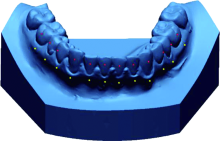
Fig.1
Define the reference point"

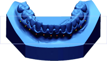
Fig.2
Establish the reference plane"

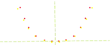
Fig.3
Establish the rectangular coordinate system"
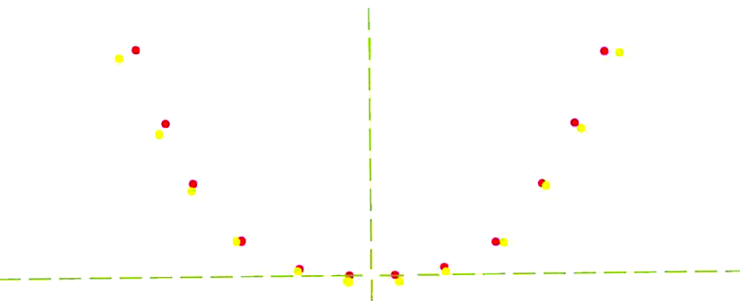
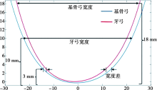
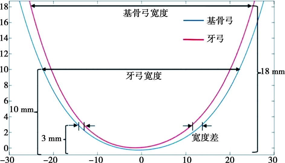
Fig.4
Measure the width and width difference"
Tab.1
Width of Skeletal Class Ⅱ and Ⅰ dental arches and basal bone x ¯±s,mm"
| 分段 | 测量部位 | Ⅱ类 | Ⅰ类 | P |
|---|---|---|---|---|
| 前段 | 牙弓 | 27.12±2.53 | 23.54±2.49 | 0.000** |
| 基骨 | 25.54±1.73 | 25.61±1.34 | 0.887 | |
| 中段 | 牙弓 | 39.06±2.09 | 38.57±1.96 | 0.461 |
| 基骨 | 40.86±1.96 | 41.49±1.85 | 0.315 | |
| 后段 | 牙弓 | 46.86±2.73 | 47.70±2.63 | 0.343 |
| 基骨 | 50.67±2.22 | 51.94±2.43 | 0.095 |
Tab.2
The width difference between the mandibular basal bone and the dental arch in Skeletal Class Ⅱ and Class Ⅰ x ¯±s,mm"
| 分段 | 宽度差 | t | P | |
|---|---|---|---|---|
| 前段 | Ⅱ类 | -1.58±2.44 | 4.527 | 0.000* |
| Ⅰ类 | 2.08±2.59 | |||
| 中段 | Ⅱ类 | 1.80±1.49 | 2.298 | 0.027* |
| Ⅰ类 | 2.92±1.54 | |||
| 后段 | Ⅱ类 | 3.80±1.42 | 1.011 | 0.318 |
| Ⅰ类 | 4.24±1.20 |
| [1] | 傅民魁, 张丁, 王邦康, 等. 中国25 392名儿童与青少年错牙合畸形患病率的调查[J]. 中华口腔医学杂志, 2002, 37(5): 371-373. |
| [2] | Ronay V, Miner RM, Will LA, et al. Mandibular arch form: The relationship between dental and basal anatomy[J]. Am J OrthodDentofacial Orthop, 2008, 134(3): 430-438. |
| [3] | Kim KY, Bayome M, Kim K, et al. Three-dimensional evaluation of the relationship between dental and basal arch forms in normal occlusion[J]. Korean J Orthod, 2011, 41(4): 288. |
| [4] | 奚祺, 吴国锋. 数字化口内扫描技术的发展与应用[J]. 实用口腔医学杂志, 2021, 37(1): 136-140. |
| [5] | Hayama K, Arai K, Ishikawa H. Correlation between upper and lower dental arch froms by fitting of fourth-order polynomials[J]. Orthod Waves, 2000, 59:303-311. |
| [6] | 高盟. 骨性Ⅱ类错牙合拔牙治疗前后基骨与牙弓形态的研究[D]. 西安: 中国人民解放军空军军医大学, 2018. |
| [7] |
Wen YF, Wong HM, Pei T, et al. Adolescent dental arch development among southern Chinese in Hong Kong: A geometric morphometric approach[J]. Sci Rep, 2019, 9(1): 18526.
doi: 10.1038/s41598-019-55073-2 pmid: 31811230 |
| [8] | 高盟, 刘冬梅, 程锦, 等. 骨性Ⅱ类错牙合拔牙矫治前后下颌牙弓与基骨形态的变化[J]. 中华医学美学美容杂志, 2018, 24(3): 188-191. |
| [9] | Brash JC. The etiology of irregularity and malocclusion of the teeth[M]. London: Dental Board of the United Kingdom, 1956. |
| [10] | Andrews L. The six elements of orofacial harmony[J]. Andrews J, 2000(1):13-22. |
| [11] | Ramón R, Adanero A, Miegimolle M. A new approach to diagnosis to posterior cross bite: Intraoral photography and walaridge[J]. Int J Environ Res Public Health, 2022, 19(15): 9443. |
| [12] | Uysal T, Badel M, Serdar U, et al. Dental and alveolar arch widths in normal occlusion, Class Ⅱ division 1 and Class Ⅱ division 2[J]. Angle Orthod, 2005, 75(6): 941-947. |
| [13] | Gupta D, Miner RM, Arai K, et al. Comparison of the mandibular dental and basal arch forms in adults and children with Class Ⅰ and Class Ⅱmalocclusions[J]. Am J Orthod Dentofacial Orthop, 2010, 138(1): 10. e1-10. e8;discussion 10-11. |
| [14] |
Ueno K, Kumabe S, Nakatsuka M, et al. Factors influencing dental arch form[J]. Okajimas Folia Anat Jpn, 2019, 96(1): 31-46.
doi: 10.2535/ofaj.96.31 pmid: 31462623 |
| [15] | 李蓉, 戴宁, 陈嵘, 等. 安氏Ⅱ1类错牙合与正常牙合牙弓宽度的研究[J]. 临床口腔医学杂志, 2009, 25(11): 677-679. |
| [16] | Buschang PH, Roldan SI, Tadlock LP. Guidelines for assessing the growth and development of orthodontic patients[J]. Semin Orthod, 2017, 23(4): 321-335. |
| [17] | 徐舒豪, 彭薇, 黄诗言, 等. 不同矢状骨性错牙合畸形牙弓及基骨宽度的比较研究[J]. 成都医学院学报, 2024, 19(1): 28-33. |
| [18] | 陈上, 厉松. 自锁托槽解除上颌牙列拥挤后牙弓与牙齿的三维变化研究[J]. 中华口腔正畸学杂志, 2016, 23(2): 67-72. |
| Viewed | ||||||
|
Full text |
|
|||||
|
Abstract |
|
|||||