
Stomatology ›› 2026, Vol. 46 ›› Issue (1): 48-53.doi: 10.13591/j.cnki.kqyx.2026.01.008
• Basic and Clinical Research • Previous Articles Next Articles
YIN Xiaoli1,2, LIU Yang3( )
)
Received:2025-06-25
Online:2026-01-28
Published:2026-01-16
Contact:
LIU Yang
E-mail:liu@scu.edu.cn
CLC Number:
YIN Xiaoli, LIU Yang. Correlation between the changes of articular disc and mandibular morphology in adult women with temporomandibular disorders symptoms[J]. Stomatology, 2026, 46(1): 48-53.
Tab.1
Cephalometric measurements and definitions"
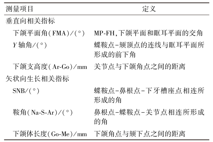 |
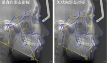
Fig.1
Cephalometric measurements used in the study"

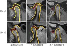
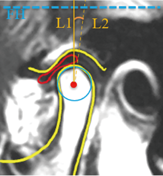
Fig.2
Diagnosis of disc displacements in MRIs"

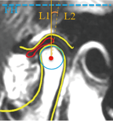
Fig.3
Evaluation criteria for disc displacements in MRIs"
Tab.2
The quantity of position and morphology of left and right discs"
| 关节盘 | 总数 | 位置 | 形态 | |||
|---|---|---|---|---|---|---|
| 正常 | DDwR | DDwoR | 基本正常 | 异常 | ||
| 右侧R | 223 | 84 | 48 | 91 | 120 | 103 |
| 左侧L | 223 | 69 | 77 | 77 | 125 | 98 |
Tab.3
Comparison of cephalometric measurements in normal, DDR and DDNR groups"
| 测量项目 | 组别 | F | P | |||||
|---|---|---|---|---|---|---|---|---|
| 正常 | DDwR | DDwoR | ||||||
| 垂直向 | FMA | R | 24.47±5.88 | 24.71±5.76 | 28.24±6.84 | 9.32 | 0.000** | |
| L | 23.83±5.92 | 25.48±5.57 | 28.64±7.03 | 11.38 | 0.000** | |||
| Y轴角 | R | 62.11±3.41 | 61.55±3.65 | 63.08±3.91 | 3.10 | 0.047* | ||
| L | 61.70±3.44 | 61.87±3.59 | 63.52±3.84 | 5.74 | 0.004** | |||
| Ar-Go | R | 46.50±4.41 | 44.82±3.75 | 43.77±4.13 | 9.45 | 0.000** | ||
| L | 46.88±4.20 | 45.24±4.04 | 43.14±3.95 | 15.67 | 0.000** | |||
| 矢状向 | SNB | R | 77.75±3.60 | 77.65±3.06 | 76.60±3.91 | 2.56 | 0.080 | |
| L | 77.69±3.57 | 77.88±3.53 | 76.24±3.67 | 4.74 | 0.010* | |||
| Na-S-Ar | R | 127.36±5.51 | 125.81±3.97 | 126.05±5.26 | 1.99 | 0.139 | ||
| L | 127.69±5.09 | 125.98±4.63 | 125.92±5.54 | 2.78 | 0.064 | |||
| Go-Me | R | 67.16±4.53 | 67.61±3.94 | 65.83±5.52 | 2.70 | 0.070 | ||
| L | 67.16±4.82 | 66.78±4.00 | 66.24±5.70 | 0.66 | 0.521 | |||
Tab.4
Pairwise comparison of the correlation between disc position and the vertical and sagittal morphology of the mandible"
| 测量项目 | P | |||
|---|---|---|---|---|
| 正常-DDwR | 正常-DDwoR | DDwR-DDwoR | ||
| FMA | R | 0.831 | 0.000** | 0.002** |
| L | 0.111 | 0.000** | 0.002** | |
| Y轴角 | R | 0.403 | 0.082 | 0.021* |
| L | 0.787 | 0.003** | 0.005** | |
| Ar-Go | R | 0.027* | 0.000** | 0.159 |
| L | 0.016* | 0.000** | 0.001** | |
| SNB | R | - | - | - |
| L | 0.751 | 0.016* | 0.005** | |
Tab.5
Comparison of cephalometric measurements in normal and abnormal groups"
| 测量项目 | 组别 | F | P | |||
|---|---|---|---|---|---|---|
| 基本正常 | 异常 | |||||
| 垂 直 向 | FMA | R | 24.76±5.59 | 27.58±7.15 | 10.92 | 0.001** |
| L | 24.33±5.72 | 28.27±6.78 | 22.17 | 0.000** | ||
| Y轴角 | R | 62.05±3.40 | 63.28±4.02 | 6.16 | 0.018* | |
| L | 61.69±3.48 | 63.27±3.81 | 10.34 | 0.001** | ||
| Ar-Go | R | 46.02±4.20 | 43.87±4.19 | 14.54 | 0.000** | |
| L | 46.36±4.14 | 43.32±3.95 | 30.68 | 0.000** | ||
| 矢 状 向 | SNB | R | 77.80±3.40 | 76.62±3.84 | 5.90 | 0.016* |
| L | 77.82±3.56 | 76.54±3.66 | 6.91 | 0.009** | ||
| Na-S-Ar | R | 126.71±5.13 | 126.24±5.16 | 0.45 | 0.504 | |
| L | 127.07±4.96 | 125.76±5.28 | 3.59 | 0.059 | ||
| Go-Me | R | 67.26±4.40 | 66.08±5.35 | 3.27 | 0.072 | |
| L | 67.27±4.44 | 66.00±5.35 | 3.76 | 0.054 | ||
| [1] |
Ohrbach R, Dworkin SF. The evolution of TMD diagnosis: Past, present, future[J]. J Dent Res, 2016, 95(10): 1093-1101.
doi: 10.1177/0022034516653922 |
| [2] |
Weinberg LA. The role of stress, occlusion, and condyle position in TMJ dysfunction-pain[J]. J Prosthet Dent, 1983, 49(4): 532-545.
pmid: 6573501 |
| [3] |
Slade GD, Bair E, Greenspan JD, et al. Signs and symptoms of first-onset TMD and sociodemographic predictors of its development: The OPPERA prospective cohort study[J]. J Pain, 2013, 14(12 Suppl): T20-32.e1-3.
doi: 10.1016/j.jpain.2013.07.014 |
| [4] |
Davant TS 6th, Greene CS, Perry HT, et al. A quantitative computer-assisted analysis of disc displacement in patients with internal derangement using sagittal view magnetic resonance imaging[J]. J Oral Maxillofac Surg, 1993, 51(9): 974-979;discussion 979-981.
doi: 10.1016/S0278-2391(10)80038-2 |
| [5] |
Haiter-Neto F, Hollender L, Barclay P, et al. Disk position and the bilaminar zone of the temporomandibular joint in asymptomatic young individuals by magnetic resonance imaging[J]. Oral Surg Oral Med Oral Pathol Oral Radiol Endod, 2002, 94(3): 372-378.
doi: 10.1067/moe.2002.127086 |
| [6] |
Katzberg RW, Westesson PL, Tallents RH, et al. Anatomic disorders of the temporomandibular joint disc in asymptomatic subjects[J]. J Oral Maxillofac Surg, 1996, 54(2): 147-153;discussion 153-155.
doi: 10.1016/S0278-2391(96)90436-X |
| [7] |
Tasaki MM, Westesson PL, Isberg AM, et al. Classification and prevalence of temporomandibular joint disk displacement in patients and symptom-free volunteers[J]. Am J Orthod Dentofacial Orthop, 1996, 109(3): 249-262.
doi: 10.1016/S0889-5406(96)70148-8 |
| [8] | Paknahad M, Shahidi S, Akhlaghian M, et al. Is mandibular Fossa morphology and articular eminence inclination associated with temporomandibular dysfunction[J]. J Dent (Shiraz), 2016, 17(2): 134-141. |
| [9] |
Gökalp H. Disc position in clinically asymptomatic, pretreatment adolescents with Class Ⅰ, Ⅱ, or Ⅲ malocclusion[J]. J Orofac Orthop / Fortschr Der Kieferorthopädie, 2016, 77(3): 194-202.
doi: 10.1007/s00056-016-0024-6 |
| [10] |
Kristensen KD, Schmidt B, Stoustrup P, et al. Idiopathic condylar resorptions: 3-dimensional condylar bony deformation, signs and symptoms[J]. Am J Orthod Dentofac Orthop, 2017, 152(2): 214-223.
doi: 10.1016/j.ajodo.2016.12.020 |
| [11] | Paknahad M, Shahidi S, Abbaszade H. Correlation between condylar position and different sagittal skeletal facial types[J]. J Orofac Orthop/Fortschr Der Kieferorthopädie, 2016, 77(5): 350-356. |
| [12] |
Shu C, Xiong X, Huang LW, et al. The relation of cephalometric features to internal derangements of the temporomandibular joint: A systematic review and meta-analysis of observational studies[J]. Orthod Craniofac Res, 2021, 24(3): 305-313.
doi: 10.1111/ocr.12454 pmid: 33290631 |
| [13] | Shi JJ, Zhang F, Zhou YQ, et al. The relationship between partial disc displacement and mandibular dysplasia in female adolescents[J]. Med Sci Monit, 2010, 16(6): CR283-CR288. |
| [14] | 刘加强, 吴勇, 孙良严, 等. 颌骨垂直向异常患者中颞下颌关节结构的情况分析[J]. 口腔颌面修复学杂志, 2015, 16(6): 335-338. |
| [15] |
Jeon DM, Jung WS, Mah SJ, et al. The effects of TMJ symptoms on skeletal morphology in orthodontic patients with TMJ disc displacement[J]. Acta Odontol Scand, 2014, 72(8): 776-782.
doi: 10.3109/00016357.2014.906650 |
| [16] |
Byun ES, Ahn SJ, Kim TW. Relationship between internal derangement of the temporomandibular joint and dentofacial morphology in women with anterior open bite[J]. Am J Orthod Dentofac Orthop, 2005, 128(1): 87-95.
doi: 10.1016/j.ajodo.2004.01.028 |
| [17] |
Ioi H, Matsumoto R, Nishioka M, et al. Relationship of TMJ osteoarthritis/osteoarthrosis to head posture and dentofacial morphology[J]. Orthod Craniofac Res, 2008, 11(1): 8-16.
doi: 10.1111/j.1601-6343.2008.00406.x pmid: 18199075 |
| [18] |
Cedströmer AL, Andlin-Sobocki A, Abbu N, et al. Condylar alterations and facial growth in children with juvenile idiopathic arthritis[J]. J Orofac Orthop / Fortschr Der Kieferorthopädie, 2020, 81(3): 163-171.
doi: 10.1007/s00056-020-00216-8 |
| [19] |
Ahn SJ, Baek SH, Kim TW, et al. Discrimination of internal derangement of temporomandibular joint by lateral cephalometric analysis[J]. Am J Orthod Dentofac Orthop, 2006, 130(3): 331-339.
doi: 10.1016/j.ajodo.2005.02.019 |
| [20] |
Taşkaya-Yilmaz N, Oğütcen-Toller M. Magnetic resonance imaging evaluation of temporomandibular joint disc deformities in relation to type of disc displacement[J]. J Oral Maxillofac Surg, 2001, 59(8): 860-865;discussion 865-866.
doi: 10.1053/joms.2001.25016 |
| [21] |
Amaral RD, Damasceno NN, de Souza LA, et al. Magnetic resonance images of patients with temporomandibular disorders: Prevalence and correlation between disk morphology and displacement[J]. Eur J Radiol, 2013, 82(6): 990-994.
doi: 10.1016/j.ejrad.2013.01.002 pmid: 23369857 |
| [22] | 马绪臣, 张震康. 颞下颌关节紊乱病双轴诊断的临床意义和规范治疗的必要性[J]. 中华口腔医学杂志, 2005, 40(5): 353-355. |
| [23] |
Weedon S, Ahmed N, Sidebottom AJ. Prospective assessment of outcomes following disposable arthroscopy of the temporomandibular joint[J]. Br J Oral Maxillofac Surg, 2013, 51(7): 625-629.
doi: 10.1016/j.bjoms.2013.06.004 pmid: 23886497 |
| [24] |
Incesu L, Taşkaya-Yilmaz N, Oğütcen-Toller M, et al. Relation-ship of condylar position to disc position and morphology[J]. Eur J Radiol, 2004, 51(3): 269-273.
pmid: 15294336 |
| [25] |
Rabelo KA, Sousa Melo SL, Torres MGG, et al. Assessment of condyle position, Fossa morphology, and disk displacement in symptomatic patients[J]. Oral Surg Oral Med Oral Pathol Oral Radiol, 2017, 124(2): 199-207.
doi: 10.1016/j.oooo.2017.04.007 |
| [26] |
Suzuki K, Mito T, Ishizaki K, et al. Mandibular lateral translation during symmetric mandibular function in relation to patterns of intracapsular derangement of TMJ[J]. Int J Stomatol Occlusion Med, 2009, 2(1): 16-23.
doi: 10.1007/s12548-009-0003-2 |
| [27] |
Bertram S, Moriggl A, Rudisch A, et al. Structural characteristics of bilateral temporomandibular joint disc displacement without reduction and osteoarthrosis are important determinants of horizontal mandibular and vertical ramus deficiency: A magnetic resonance imaging study[J]. J Oral Maxillofac Surg, 2011, 69(7): 1898-1904.
doi: 10.1016/j.joms.2010.12.026 |
| [28] |
Hsieh YJ, Darvann TA, Hermann NV, et al. Three-dimensional assessment of facial morphology in children and adolescents with juvenile idiopathic arthritis and moderate to severe TMJ involvement using 3D surface scans[J]. Clin Oral Investig, 2020, 24(2): 799-807.
doi: 10.1007/s00784-019-02962-5 |
| [29] |
Lin M, Xu YF, Wu H, et al. Comparative cone-beam computed tomography evaluation of temporomandibular joint position and morphology in female patients with skeletal Class Ⅱ malocclusion[J]. J Int Med Res, 2020, 48(2): 300060519892388.
doi: 10.1177/0300060519892388 |
| [30] |
Noh KJ, Baik HS, Han SS, et al. Differences in mandibular condyle and glenoid Fossa morphology in relation to vertical and sagittal skeletal patterns: A cone-beam computed tomography study[J]. Korean J Orthod, 2021, 51(2): 126-134.
doi: 10.4041/kjod.2021.51.2.126 pmid: 33678628 |
| [31] | Ma HD, Teng HD, Li AN, et al. The pressure in the temporomandibular joint in the patients with maxillofacial deformities[J]. J Stomatol Oral Maxillofac Surg, 2023, 124(1S): 101285. |
| [32] |
Wang J, Dong X, Lei J, et al. β-catenin orchestrates Gli1+ cell fate in condylar development and TMJOA[J]. J Dent Res, 2024, 103(12): 1291-1301.
doi: 10.1177/00220345241274354 |
| [33] |
Li W, Wu N, Wang JM, et al. Role of HIF-2α/NF-κB pathway in mechanical stress-induced temporomandibular joint osteoarthritis[J]. Oral Dis, 2022, 28(8): 2239-2247.
doi: 10.1111/odi.v28.8 |
| [34] |
Arieta-Miranda JM, Silva-Valencia M, Flores-Mir C, et al. Spatial analysis of condyle position according to sagittal skeletal relationship, assessed by cone beam computed tomography[J]. Prog Orthod, 2013, 14: 36.
doi: 10.1186/2196-1042-14-36 pmid: 24325842 |
| [35] |
Park IY, Kim JH, Park YH. Three-dimensional cone-beam computed tomography based comparison of condylar position and morphology according to the vertical skeletal pattern[J]. Korean J Orthod, 2015, 45(2): 66-73.
doi: 10.4041/kjod.2015.45.2.66 |
| [36] |
Saccomanno S, Deli R, DI Cintio G, et al. Retrospective epidemiological study of mandibular rotational types in patients with orthodontical malocclusion[J]. Acta Otorhinolaryngol Ital, 2018, 38(2): 160-165.
doi: 10.14639/0392-100X-1682 pmid: 29967561 |
| [37] |
Jung WS, Kim H, Jeon DM, et al. Magnetic resonance imaging-verified temporomandibular joint disk displacement in relation to sagittal and vertical jaw deformities[J]. Int J Oral Maxillofac Surg, 2013, 42(9): 1108-1115.
doi: 10.1016/j.ijom.2013.03.012 |
| Viewed | ||||||
|
Full text |
|
|||||
|
Abstract |
|
|||||