
Stomatology ›› 2023, Vol. 43 ›› Issue (7): 638-642.doi: 10.13591/j.cnki.kqyx.2023.07.010
• Clinical Research • Previous Articles Next Articles
FAN Siyu,XUE Yiwen,SONG Xiao,DENG Runzhi,HAO Jing( )
)
Revised:2023-02-01
Online:2023-07-28
Published:2023-07-28
CLC Number:
FAN Siyu, XUE Yiwen, SONG Xiao, DENG Runzhi, HAO Jing. Study on clinical characteristics of impacted maxillary canine-linked incisor root resorption[J]. Stomatology, 2023, 43(7): 638-642.

Fig.1
The angle and distance to MP"
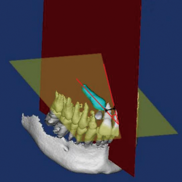
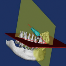
Fig.2
The angle and distance to OP"

Tab.1
Characteristics of patients with impacted canine"
| 测量项目 | 组别 | χ2 | P | ||
|---|---|---|---|---|---|
| 11~14岁 | 15~19岁 | 20~30岁 | |||
| 性别 | |||||
| 男 | 5 | 5 | 5 | 0.38 | 0.83 |
| 女 | 14 | 10 | 9 | ||
| 安氏分类 | |||||
| Ⅰ类 | 3 | 1 | 1 | 5.10 | 0.28 |
| Ⅱ类 | 13 | 13 | 8 | ||
| Ⅲ类 | 3 | 1 | 5 | ||
| 覆?? | |||||
| 正常 | 1 | 3 | 7 | 9.42 | 0.01* |
| 异常 | 18 | 12 | 7 | ||
| 覆盖 | |||||
| 正常 | 4 | 7 | 11 | 10.75 | 0.01* |
| 异常 | 15 | 8 | 3 | ||
| 侧切牙形态 | |||||
| 正常 | 14 | 13 | 6 | 6.82 | 0.03* |
| 异常 | 5 | 2 | 8 | ||
| 乳牙滞留 | |||||
| 是 | 10 | 6 | 4 | 1.95 | 0.38 |
| 否 | 9 | 9 | 10 | ||
| 阻生侧牙列间隙 | |||||
| 是 | 6 | 1 | 7 | 7.49 | 0.02* |
| 否 | 12 | 14 | 7 | ||
Tab.2
Characteristics of impacted canine and adjacent teeth"
| 测量项目 | 组别 | χ2 | P | ||
|---|---|---|---|---|---|
| 11~14岁 | 15~19岁 | 20~30岁 | |||
| 阻生牙位置 | |||||
| 唇侧 | 12 | 9 | 2 | 8.99 | 0.01* |
| 腭侧 | 7 | 6 | 12 | ||
| 牙根吸收牙位 | |||||
| 中切牙 | 6 | 3 | 4 | 11.22 | 0.02* |
| 侧切牙 | 12 | 7 | 3 | ||
| 共吸收 | 1 | 5 | 7 | ||
| 牙根吸收程度 | |||||
| 轻度 | 4 | 2 | 6 | 3.36 | 0.50 |
| 中度 | 7 | 6 | 4 | ||
| 重度 | 9 | 12 | 11 | ||
| 邻牙接触 | |||||
| 是 | 18 | 11 | 9 | 7.72 | 0.13 |
| 否 | 1 | 4 | 5 | ||
| 牙囊存在 | |||||
| 是 | 18 | 14 | 13 | 0.06 | 0.97 |
| 否 | 1 | 1 | 1 | ||
| 牙囊增大 | |||||
| 是 | 5 | 4 | 1 | 2.62 | 0.27 |
| 否 | 14 | 11 | 13 | ||
Tab.3
Angle, distance and arch width"
| 测量项目 | 组别 | P | ||
|---|---|---|---|---|
| 11~14岁 | 15~19岁 | 20~30岁 | ||
| 阻生尖牙与正中矢状面的角度/(°) | 41.43±7.61 | 42.72±12.94 | 53.70±8.76 | 0.14 |
| 阻生尖牙与正中矢状面的距离/mm | 4.54±1.68 | 4.53±1.50 | 2.53±0.95 | 0.09 |
| 阻生尖牙与咬合平面的角度/(°) | 45.66±8.81 | 42.12±8.33 | 45.04±10.78 | 0.83 |
| 阻生尖牙与咬合平面的距离/mm | 10.73±1.53 | 12.64±1.80 | 10.48±2.26 | 0.17 |
| 前牙弓宽度/mm | 36.03±1.23 | 33.79±1.48 | 36.43±1.33 | 0.01* |
| 后牙弓宽度/mm | 47.41±1.97 | 47.11±2.11 | 47.81±1.27 | 0.82 |
| [1] | Pant BD, Rajbhandari A, Pradhan R, et al. Impacted canine in orthodontic patients of a tertiary care hospital: A descriptive cross-sectional study[J]. J Nepal Med Assoc, 2021, 59(244):1215-1218. |
| [2] |
Lövgren ML, Dahl O, Uribe P, et al. Prevalence of impacted maxillary canines-an epidemiological study in a region with systematically implemented interceptive treatment[J]. Eur J Orthod, 2019, 41(5):454-459.
doi: 10.1093/ejo/cjz056 |
| [3] |
Sajnani AK, King NM. Complications associated with the occurrence and treatment of impacted maxillary canines[J]. Singapore Dent J, 2014, 35:53-57.
doi: 10.1016/j.sdj.2014.07.001 pmid: 25496586 |
| [4] |
Sarica I, Derindag G, Kurtuldu E, et al. A retrospective study: Do all impacted teeth cause pathology?[J]. Niger J Clin Pract, 2019, 22(4):527-533.
doi: 10.4103/njcp.njcp_563_18 pmid: 30975958 |
| [5] |
Ghaffar F, Sukhia RH, Fida M. Association between maxillary transverse discrepancy and occurrence of potentially impacted maxillary canines in mixed dentition patients[J]. Int Orthod, 2019, 17(3):554-561.
doi: 10.1016/j.ortho.2019.06.016 |
| [6] |
Cacciatore G, Poletti L, Sforza C. Early diagnosed impacted maxillary canines and the morphology of the maxilla: A three-dimensional study[J]. Prog Orthod, 2018, 19(1):20.
doi: 10.1186/s40510-018-0220-6 pmid: 30009340 |
| [7] |
Wang H, Li T, Lv C, et al. Risk factors for maxillary impacted canine-linked severe lateral incisor root resorption: A cone-beam computed tomography study[J]. Am J Orthod Dentofacial Orthop, 2020, 158(3):410-419.
doi: 10.1016/j.ajodo.2019.09.015 |
| [8] |
Dağsuyu İM, Kahraman F, Okşayan R. Three-dimensional evaluation of angular, linear, and resorption features of maxillary impacted canines on cone-beam computed tomography[J]. Oral Radiol, 2018, 34(1):66-72.
doi: 10.1007/s11282-017-0289-5 pmid: 30484094 |
| [9] |
Hajeer MY, Al-Homsi HK, Alfailany DT, et al. Evaluation of the diagnostic accuracy of CBCT-based interpretations of maxillary impacted canines compared to those of conventional radiography: An in vitro study[J]. Int Orthod, 2022, 20(2):100639.
doi: 10.1016/j.ortho.2022.100639 |
| [10] |
Lai CS, Suter VGA, Katsaros C, et al. Localization of impacted maxillary canines and root resorption of neighbouring teeth: A study assessing the diagnostic value of panoramic radiographs in two groups of observers[J]. Eur J Orthod, 2014, 36(4):450-456.
doi: 10.1093/ejo/cjt074 |
| [11] |
Wriedt S, Jaklin J, Al-Nawas B, et al. Impacted upper canines: Examination and treatment proposal based on 3D versus 2D diagnosis[J]. J Orofac Orthop, 2012, 73(1):28-40.
doi: 10.1007/s00056-011-0058-8 pmid: 22246048 |
| [12] |
Alqerban A, Jacobs R, Fieuws S, et al. Comparison of two cone beam computed tomographic systems versus panoramic imaging for localization of impacted maxillary canines and detection of root resorption[J]. Eur J Orthod, 2011, 33(1):93-102.
doi: 10.1093/ejo/cjq034 |
| [13] | Enlow DH. Handbook of facial growth[M]. 2nd ed. Philadelphia: WB Saunders, 1982:182-183. |
| [14] |
Kalavritinos M, Benetou V, Bitsanis E, et al. Incidence of incisor root resorption associated with the position of the impacted maxillary canines: A cone-beam computed tomographic study[J]. Am J Orthod Dentofacial Orthop, 2020, 157(1):73-79.
doi: 10.1016/j.ajodo.2019.02.016 |
| [15] | Guarnieri R, Cavallini C, Vernucci R, et al. Impacted maxillary canines and root resorption of adjacent teeth: A retrospective observational study[J]. Med Oral Patol Oral Cir Bucal, 2016, 21(6):e743-e750. |
| [16] |
Chaushu S, Kaczor-Urbanowicz K, Zadurska M, et al. Predisposing factors for severe incisor root resorption associated with impacted maxillary canines[J]. Am J Orthod Dentofacial Orthop, 2015, 147(1):52-60.
doi: 10.1016/j.ajodo.2014.09.012 |
| [17] | 田玉楼, 朴美玲, 赵震锦, 等. 上颌前牙埋伏阻生与矢状骨面型的关系[J]. 上海口腔医学, 2014, 23(1):99-102. |
| [18] |
Sajnani AK, King NM. Dental anomalies associated with buccally- and palatally-impacted maxillary canines[J]. J Investig Clin Dent, 2014, 5(3):208-213.
doi: 10.1111/jicd.12035 |
| [19] |
Kim Y, Hyun HK, Jang KT. Morphological relationship analysis of impacted maxillary canines and the adjacent teeth on 3-dimensional reconstructed CT images[J]. Angle Orthod, 2017, 87(4):590-597.
doi: 10.2319/071516-554.1 pmid: 28156127 |
| [20] |
Yan B, Sun ZY, Fields H, et al. Etiologic factors for buccal and palatal maxillary canine impaction: A perspective based on cone-beam computed tomography analyses[J]. Am J Orthod Dentofacial Orthop, 2013, 143(4):527-534.
doi: 10.1016/j.ajodo.2012.11.021 |
| [21] |
Kim Y, Hyun HK, Jang KT. The position of maxillary canine impactions and the influenced factors to adjacent root resorption in the Korean population[J]. Eur J Orthod, 2012, 34(3):302-306.
doi: 10.1093/ejo/cjr002 |
| [22] | Ali IH, Al-Turaihi BA, Mohammed LK, et al. Root resorption of teeth adjacent to untreated impacted maxillary canines: A CBCT study[J]. Biomed Res Int, 2021, 2021:6635575. |
| [23] |
Jung YH, Liang H, Benson BW, et al. The assessment of impacted maxillary canine position with panoramic radiography and cone beam CT[J]. Dentomaxillofac Radiol, 2012, 41(5):356-360.
doi: 10.1259/dmfr/14055036 |
| [24] |
Kim SH, Son WS, Yamaguchi T, et al. Assessment of the root apex position of impacted maxillary canines on panoramic films[J]. Am J Orthod Dentofacial Orthop, 2017, 152(4):489-493.
doi: 10.1016/j.ajodo.2017.01.027 |
| [25] |
Al-Kyssi HA, Al-Mogahed NM, Altawili ZM, et al. Predictive factors associated with adjacent teeth root resorption of palatally impacted canines in Arabian population: A cone-beam computed tomography analysis[J]. BMC Oral Health, 2022, 22(1):220.
doi: 10.1186/s12903-022-02249-4 pmid: 35658855 |
| [26] |
Strbac GD, Foltin A, Gahleitner A, et al. The prevalence of root resorption of maxillary incisors caused by impacted maxillary canines[J]. Clin Oral Investig, 2013, 17(2):553-564.
doi: 10.1007/s00784-012-0738-9 |
| Viewed | ||||||
|
Full text |
|
|||||
|
Abstract |
|
|||||