
Stomatology ›› 2026, Vol. 46 ›› Issue (4): 289-294.doi: 10.13591/j.cnki.kqyx.2026.04.009
• Basic and Clinical Research • Previous Articles Next Articles
ZHAO Zhuangzhuang1,2,3, ZHANG Ping1,2,3, JIANG Hongbing1,2,3, YAN Enshi2,3,4( ), XU Rongyao1,2,3()
Received:2025-11-06
Online:2026-04-28
Published:2026-04-17
CLC Number:
ZHAO Zhuangzhuang, ZHANG Ping, JIANG Hongbing, YAN Enshi, XU Rongyao. MRI analysis of preoperative and postoperative changes of masticatory muscle morphology in unilateral anterior disc displacement of temporomandibular joint[J]. Stomatology, 2026, 46(4): 289-294.
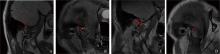
Fig.1
Diagnosis of anterior displacement of the temporomandibular joint disc"
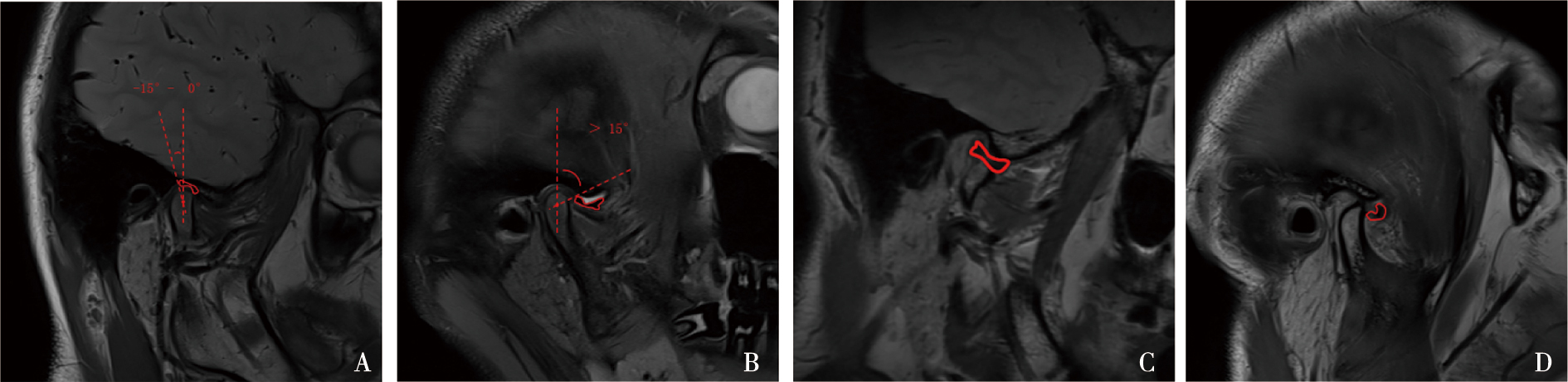
Fig.2
Height, width, thickness, and volume of the masseter muscle"
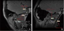
Fig.3
Height and length of the superior and inferior heads of the lateral pterygoid muscle"
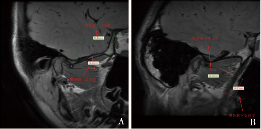

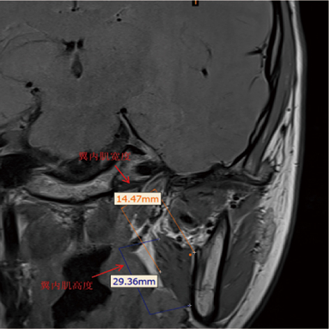
Fig.4
Height and width of the medial pterygoid muscle"
Tab. 1
Comparative analysis of masticatory muscle structures in patients with unilateral ADDwoR"
| 肌肉参数 | 不可复性患侧 | 不可复性健侧 | t | P | 患侧/健侧 |
|---|---|---|---|---|---|
| 咬肌高度/mm | 63.23±3.21 | 64.87±3.50 | -3.050 | 0.003 | 0.97 |
| 咬肌宽度/mm | 35.62±2.53 | 37.03±2.78 | -3.313 | 0.001 | 0.96 |
| 咬肌厚度/mm | 15.48±1.31 | 16.15±1.45 | -3.028 | 0.003 | 0.96 |
| 咬肌体积/cm3 | 5.73±1.84 | 7.24±2.03 | -4.867 | <0.001 | 0.79 |
| 翼外肌上头高度/mm | 7.12±1.65 | 6.22±1.58 | 3.479 | 0.001 | 1.14 |
| 翼外肌上头长度/mm | 25.67±2.43 | 24.57±2.29 | 2.910 | 0.004 | 1.04 |
| 翼外肌下头高度/mm | 14.00±1.56 | 13.62±1.62 | 1.492 | 0.138 | 1.03 |
| 翼外肌下头长度/mm | 26.23±2.19 | 26.19±2.08 | 0.117 | 0.907 | 1.00 |
| 翼内肌宽度/mm | 16.02±1.04 | 16.33±1.25 | -1.684 | 0.094 | 0.98 |
| 翼内肌高度/mm | 40.32±2.59 | 40.56±3.45 | -0.491 | 0.624 | 0.99 |
Tab.2
Comparative analysis of masticatory muscle structures in patients with unilateral ADDwR"
| 肌肉参数 | 可复性患侧 | 可复性健侧 | t | P | 患侧/健侧 |
|---|---|---|---|---|---|
| 咬肌高度/mm | 63.21±4.01 | 64.31±4.12 | -0.765 | 0.450 | 0.98 |
| 咬肌宽度/mm | 35.87±3.50 | 36.65±3.68 | -0.614 | 0.544 | 0.98 |
| 咬肌厚度/mm | 15.87±1.82 | 16.65±1.96 | -1.166 | 0.253 | 0.95 |
| 咬肌体积/cm3 | 6.45±3.29 | 7.08±3.10 | -0.557 | 0.581 | 0.91 |
| 翼外肌上头高度/mm | 7.12±0.92 | 7.43±1.07 | -0.879 | 0.387 | 0.96 |
| 翼外肌上头长度/mm | 26.02±2.11 | 24.89±2.08 | 1.526 | 0.138 | 1.05 |
| 翼外肌下头高度/mm | 12.93±1.31 | 12.38±1.25 | 1.215 | 0.234 | 1.04 |
| 翼外肌下头长度/mm | 25.54±2.59 | 25.03±2.30 | 0.589 | 0.560 | 1.02 |
| 翼内肌宽度/mm | 16.32±1.56 | 16.24±1.49 | 0.148 | 0.883 | 1.01 |
| 翼内肌高度/mm | 41.07±3.20 | 41.34±2.13 | -0.281 | 0.781 | 0.99 |
Tab.3
Comparison of masticatory muscle indices after temporomandibular joint disc repositioning surgery"
| 肌肉参数 | 不可复性患侧 | 不可复性健侧 | t | P | 患侧/健侧 |
|---|---|---|---|---|---|
| 咬肌高度/mm | 64.23±1.12 | 64.54±1.08 | -1.760 | 0.080 | 1.00 |
| 咬肌宽度/mm | 35.87±1.25 | 36.74±1.20 | -4.434 | <0.001 | 0.98 |
| 咬肌厚度/mm | 16.02±0.98 | 16.34±1.05 | -1.968 | 0.051 | 0.98 |
| 咬肌体积/cm3 | 26.87±2.15 | 26.54±2.05 | 0.981 | 0.328 | 1.01 |
| 翼外肌上头高度/mm | 6.95±0.68 | 7.32±0.72 | -3.300 | 0.001 | 0.95 |
| 翼外肌上头长度/mm | 25.69±1.10 | 25.45±1.08 | 1.375 | 0.171 | 1.00 |
| 翼外肌下头高度/mm | 13.06±0.95 | 13.45±1.02 | -2.471 | 0.015 | 0.97 |
| 翼外肌下头长度/mm | 26.24±1.12 | 25.78±1.15 | 2.531 | 0.012 | 1.02 |
| 翼内肌宽度/mm | 16.32±0.85 | 16.24±0.90 | 0.571 | 0.569 | 1.00 |
| 翼内肌高度/mm | 40.56±3.89 | 40.10±2.98 | 0.829 | 0.408 | 1.01 |
| [1] |
Tanaka E, del Pozo R, Tanaka M, et al. Three-dimensional finite element analysis of human temporomandibular joint with and without disc displacement during jaw opening[J]. Med Eng Phys, 2004, 26(6): 503-511.
doi: 10.1016/j.medengphy.2004.03.001 pmid: 15234686 |
| [2] | Rigon M, Pereira LM, Bortoluzzi MC, et al. WITHDRAWN: Arthroscopy for temporomandibular disorders[J]. Cochrane Database Syst Rev, 2015, 2015(12): CD006385. |
| [3] |
List T, Jensen RH. Temporomandibular disorders: Old ideas and new concepts[J]. Cephalalgia, 2017, 37(7): 692-704.
doi: 10.1177/0333102416686302 pmid: 28068790 |
| [4] |
Pérez del Palomar A, Doblaré M. Influence of unilateral disc displacement on the stress response of the temporomandibular joint discs during opening and mastication[J]. J Anat, 2007, 211(4): 453-463.
doi: 10.1111/j.1469-7580.2007.00796.x pmid: 17725577 |
| [5] |
Mehra P, Wolford LM. The Mitek mini anchor for TMJ disc repositioning: Surgical technique and results[J]. Int J Oral Maxillofac Surg, 2001, 30(6): 497-503.
doi: 10.1054/ijom.2001.0163 |
| [6] |
Kandasamy S, Greene CS. The evolution of temporomandibular disorders: A shift from experience to evidence[J]. J Oral Pathology Medicine, 2020, 49(6): 461-469.
doi: 10.1111/jop.v49.6 |
| [7] | Alqhtani N, AliAlshadwi A, Al-Zahrani A, et al. The role of the lateral pterygoid muscle in articular disc displacement: A cross-sectional magnetic resonance imaging study[J]. Curr Med Imaging, 2022, 18(7): 787-795. |
| [8] |
Kaneda M, Shimpo Y, Yoshida K, et al. Structural features of the temporomandibular joint evaluated by MRI and their association with oral function and craniofacial morphology in female patients with malocclusion: A cross-sectional study[J]. J Clin Med, 2025, 14(14): 4921.
doi: 10.3390/jcm14144921 |
| [9] |
Szyszka-Sommerfeld L, Sycińska-Dziarnowska M, Spagnuolo G, et al. Surface electromyography in the assessment of masticatory muscle activity in patients with pain-related temporomandibular disorders: A systematic review[J]. Front Neurol, 2023, 14: 1184036.
doi: 10.3389/fneur.2023.1184036 |
| [10] |
Saini RS, Ibrahim M, Khader MA, et al. The role of physiotherapy interventions in the management of temporomandibular joint ankylosis: A systematic review and meta-analysis[J]. Head Face Med, 2024, 20(1): 15.
doi: 10.1186/s13005-024-00416-2 |
| [11] |
Schiffman E, Ohrbach R, Truelove E, et al. Diagnostic criteria for temporomandibular disorders (DC/TMD) for clinical and research applications: Recommendations of the international RDC/TMD consortium network and orofacial pain special interest group[J]. J Oral Facial Pain Headache, 2014, 28(1): 6-27.
doi: 10.11607/jop.1151 |
| [12] | 章智宇, 杨娇艳, 邢一鸣, 等. 颞下颌关节盘复位缝合术后髁突骨再生的MRI表现及影响骨再生相关因素分析[J]. 中华口腔医学杂志, 2023, 58(10): 1004-1009. |
| [13] |
王浩, 王伟, 李强, 等. 颞下颌关节盘复位锚固术不同入路及前附着松解方式的临床效果比较[J]. 口腔疾病防治, 2025, 33(2): 129-136.
doi: 10.12016/j.issn.2096-1456.202440326 |
| [14] |
Zhou Q, Zhu HM, He DM, et al. Modified temporomandibular joint disc repositioning with mini-screw anchor: Part Ⅱ-stability evaluation by magnetic resonance imaging[J]. J Oral Maxillofac Surg, 2019, 77(2): 273-279.
doi: 10.1016/j.joms.2018.07.016 |
| [15] | 傅开元, 胡敏, 余强, 等. 颞下颌关节常规MRI检查规范及关节盘移位诊断标准的专家共识[J]. 中华口腔医学杂志, 2020, 55(9): 608-612. |
| [16] | Ghadimi N, Mehralizadeh S, Rahimian E, et al. Correlation between the masticatory muscle dimensions and internal derangement of temporomandibular joints based on magnetic resonance imaging[J]. Iran J Radiol, 2023, 20(1): e131641. |
| [17] |
Muraoka H, Kaneda T, Hirahara N, et al. Magnetic resonance image texture analysis of the lateral pterygoid muscle in patients with rheumatoid arthritis: A preliminary report[J]. Oral Radiol, 2023, 39(2): 242-247.
doi: 10.1007/s11282-022-00625-y |
| [18] | 朱妍, 李敏涵, 何佳颖, 等. 单侧颞下颌关节前移位患者咀嚼肌肌电活动与盘移位的关系[J]. 中国口腔颌面外科杂志, 2025, 23(5): 457-462. |
| [19] |
Aristokli N, Polycarpou I, Themistocleous SC, et al. Comparison of the diagnostic performance of Magnetic Resonance Imaging (MRI), ultrasound and mammography for detection of breast cancer based on tumor type, breast density and patient’s history: A review[J]. Radiography, 2022, 28(3): 848-856.
doi: 10.1016/j.radi.2022.01.006 |
| [20] |
Cezairli B, Halat İB, Balaban E. Evaluation of the correlation between articular disc displacement and the lateral pterygoid muscle using magnetic resonance imaging[J]. BMC Oral Health, 2025, 25(1): 1733.
doi: 10.1186/s12903-025-07137-1 |
| [21] |
Öçbe M, Medişoğlu MS. Magnetic resonance imaging of submental and masticatory muscle morphology and its relationship with temporomandibular joint structures[J]. Diagnostics, 2025, 15(12): 1535.
doi: 10.3390/diagnostics15121535 |
| [22] | Didier HA, Cappellari AM, Di Berardino F, et al. Electrophysiological study of the masticatory muscle activity in patients with temporomandibular disorders with and without tinnitus[J]. Clin Exp Dent Res, 2025, 11(4): e70172. |
| [23] | 魏丽丽, 李波, 程勇. 颞下颌关节紊乱病的MRI临床应用进展[J]. 口腔医学, 2024, 44(1):11-15. |
| [24] |
Kalladka M, Shastry SP, Thondebhavi M, et al. Nerve blocks in the management of acute temporomandibular disorder emergencies: A narrative review[J]. J Oral Maxillofac Anesth, 2022, 1: 15.
doi: 10.21037/joma |
| [25] |
林宇翔, 李晨曦, 龚忠诚. 颞下颌关节盘锚固术对咀嚼肌表面肌电特征影响的临床研究[J]. 口腔颌面外科杂志, 2025, 35(3): 199-204.
doi: 10.12439/kqhm.1005-4979.2025.03.005 |
| [26] |
Taşkaya-Yilmaz N, Oğütcen-Toller M. Magnetic resonance imaging evaluation of temporomandibular joint disc deformities in relation to type of disc displacement[J]. J Oral Maxillofac Surg, 2001, 59(8): 860-865;discussion865-866.
doi: 10.1053/joms.2001.25015 |
| [27] |
Zhu YK, Xu JQ, Zhang J, et al. Exercise therapy in postoperative patients with temporomandibular joint internal derangement: A systematic review[J]. J Oral Rehabil, 2024, 51(10): 2158-2168.
doi: 10.1111/joor.13780 pmid: 38873746 |
| Viewed | ||||||
|
Full text |
|
|||||
|
Abstract |
|
|||||