
Stomatology ›› 2026, Vol. 46 ›› Issue (6): 427-434.doi: 10.13591/j.cnki.kqyx.2026.06.005
• Basic and Clinical Research • Previous Articles Next Articles
WANG Mingxi1,2,3, GAO Shan1,2,3, LI Guoqing1,2,3( ), TANG Chunbo1,2,3()
Received:2025-12-24
Online:2026-06-28
Published:2026-06-17
CLC Number:
WANG Mingxi, GAO Shan, LI Guoqing, TANG Chunbo. Regulatory effect of transcription factor TBX1 on osteogenic differentiation of human dental pulp stem cells[J]. Stomatology, 2026, 46(6): 427-434.
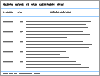
Tab.1
The primers used for RT-qPCR"
| 基因 | 引物序列(5'-3') |
|---|---|
| TBX1 | F:GCTGTGGGACGAGTTCAATC R:ACGTGGGGAACATTCGTCT |
| COL1 | F:GCTACTACCGGGCTGATGAT R:ATGTTCTCGATCTGCTGGCT |
| OSX | F:CCCACCTCAGGCTATGCTAA R:CATGGATGCCTGCCTTGTAC |
| RUNX2 | F:TGAACTCTGCACCAAGTCCT R:GGGTGGTAGAGTGGATGGAC |
| GAPDH | F:TCGTGGAAGGACTCATGACC R:ATGATGTTCTGGAGAGCCCC |
| VEGFA | F:GTGAGGTTTGATCCGCATGAT R:GACCCTGGCTTTACTGCTGTA |
| ANG1 | F:CTACCAACAACAACAGCATCC R:CTCCCTTTAGCAAAACACCTTC |
| PDGFB | F:ATGAAATGCTGAGCGACCAC G:TCCCTCGAGATGAGCTTTCC |
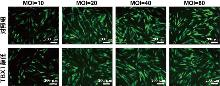
Fig.1
Representative images of hDPSCs under different MOI conditions"
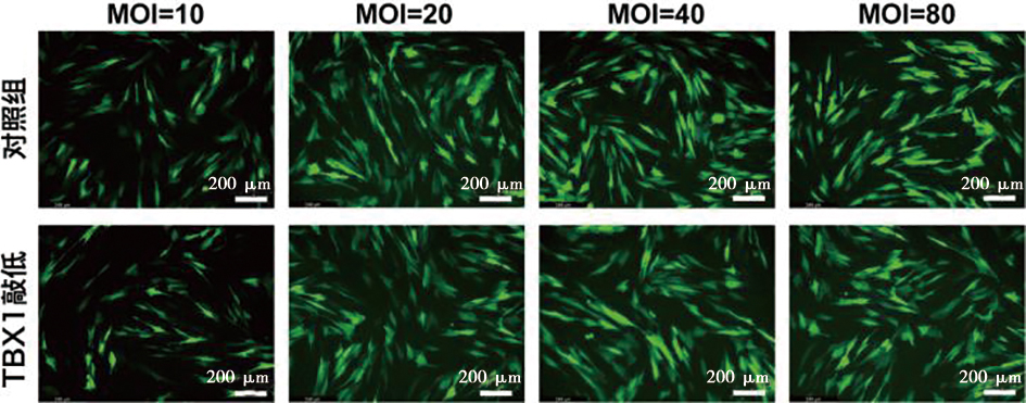
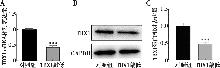
Fig.2
Validation of TBX1 knockdown efficiency"
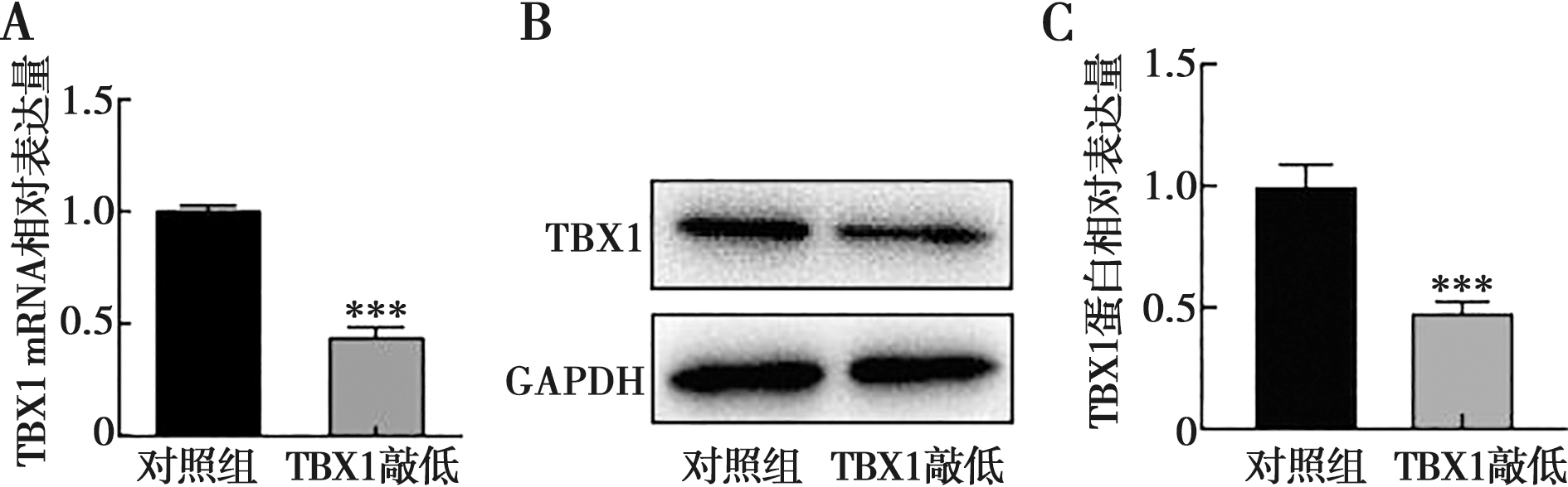
Fig.3
Knockdown of TBX1 enhances the osteogenic differentiation potential of hDPSCs in vitro"
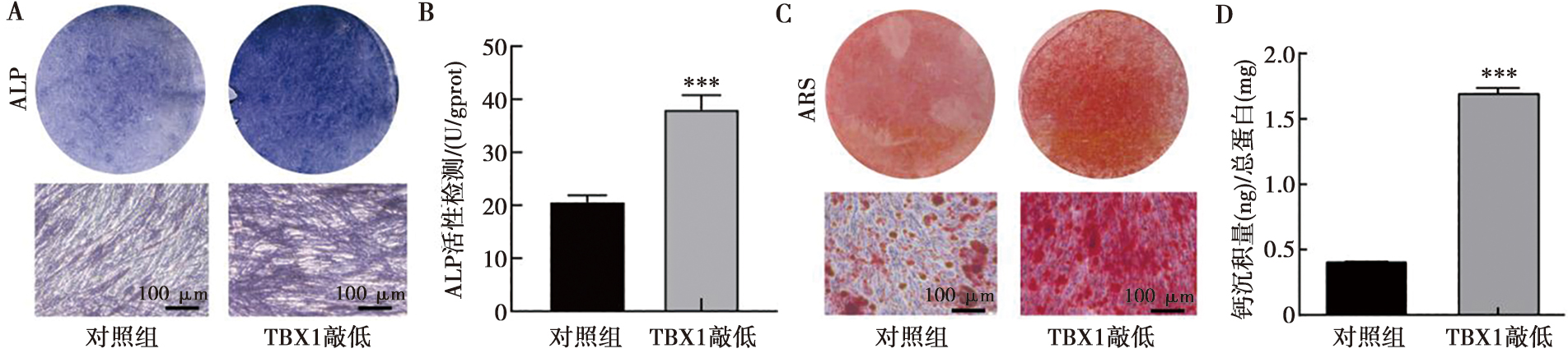
Fig.4
Knockdown of TBX1 upregulates the mRNA expression of osteogenic markers"
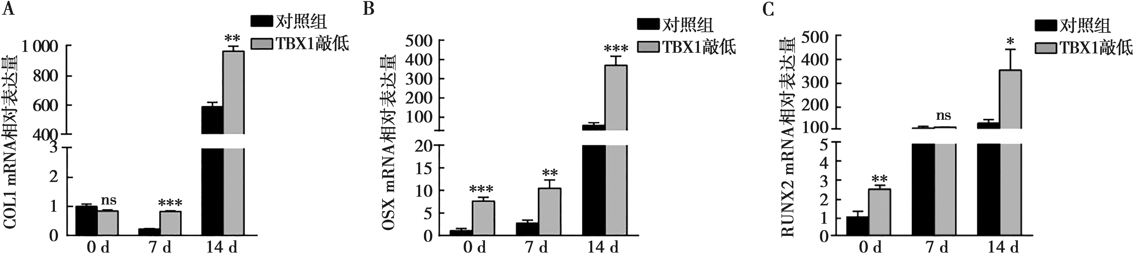
Fig.5
Knockdown of TBX1 upregulates the protein expression of osteogenic markers"
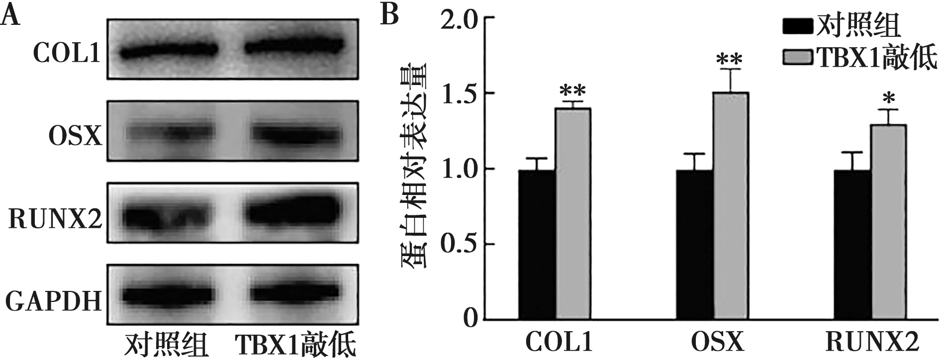
Fig.6
Knockdown of TBX1 upregulates the mRNA expression of angiogenesis-related factors"
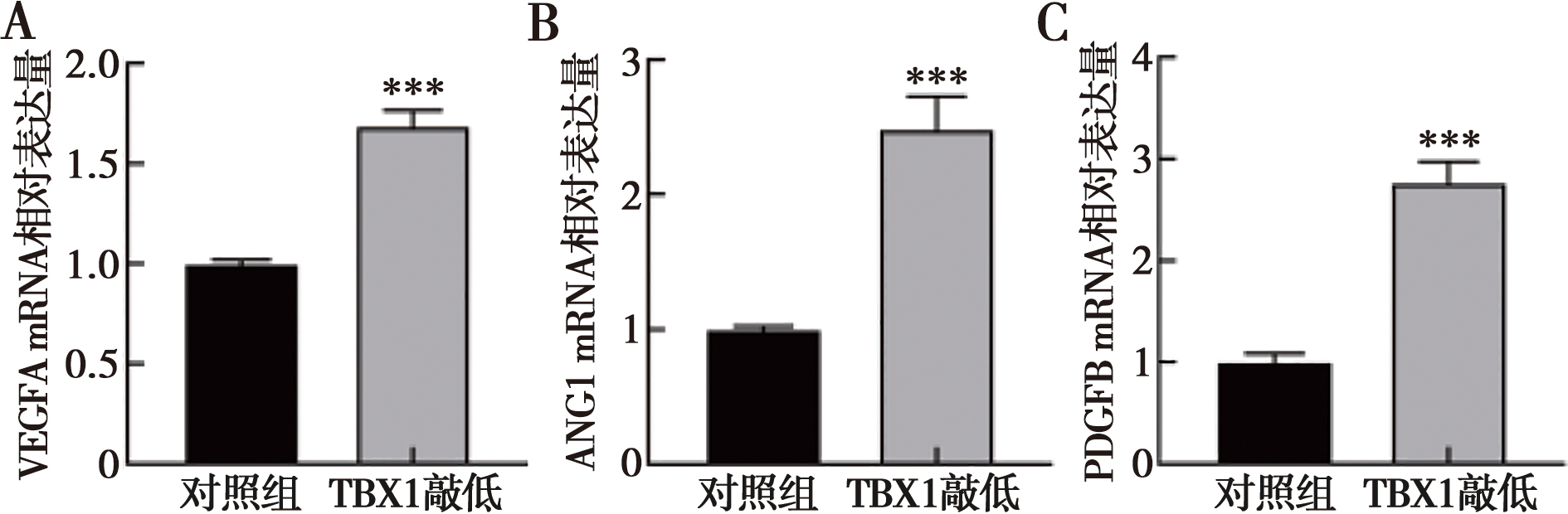
Fig.7
Knockdown of TBX1 promotes the tube formation capacity of HUVECs"
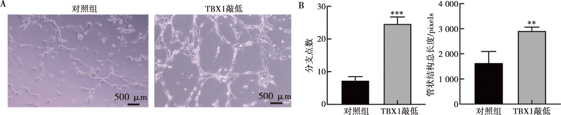
Fig.8
Knockdown of TBX1 promotes ectopic osteogenesis of hDPSCs in vivo"
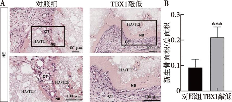
Fig.9
Knockdown of TBX1 upregulates the expression of osteogenic-related proteins in vivo"
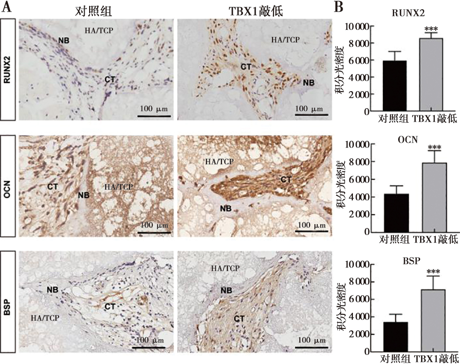
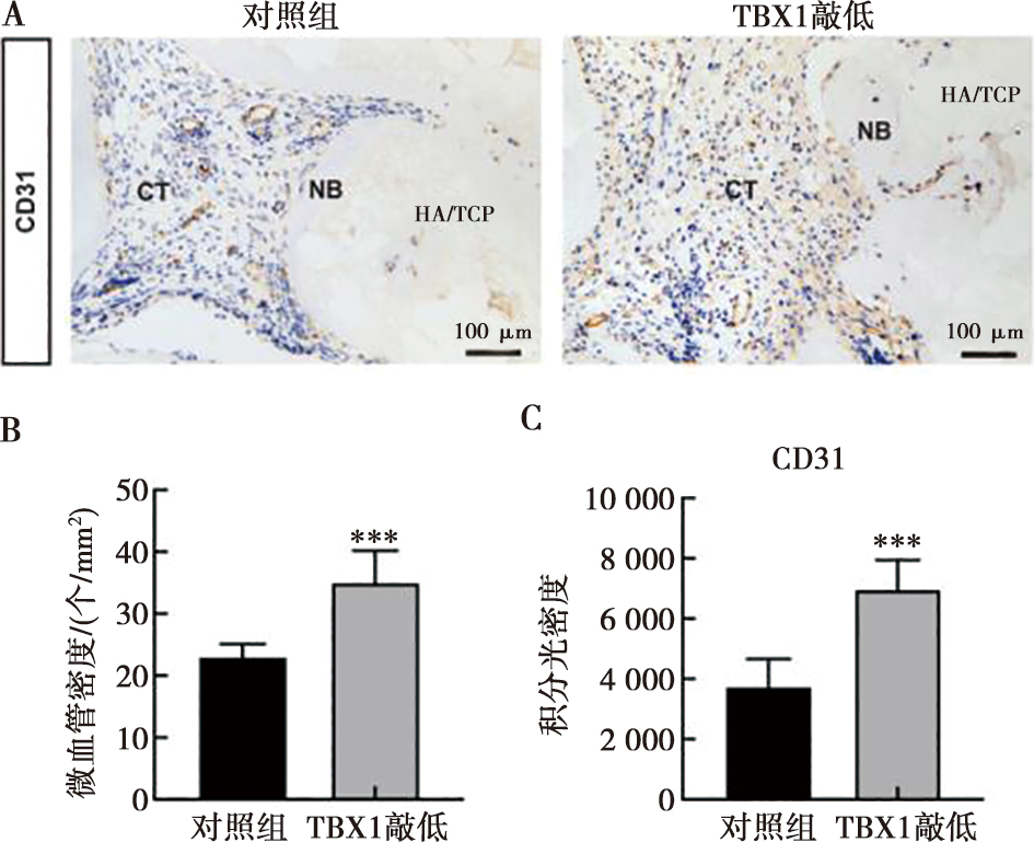
Fig.10
Knockdown of TBX1 promotes angiogenesis in vivo"
| [1] |
Raymond Y, Pastorino D, Ginebreda I, et al. Computed tomography and histological evaluation of xenogenic and biomimetic bone grafts in three-wall alveolar defects in minipigs[J]. Clin Oral Investig, 2021, 25(12): 6695-6706.
doi: 10.1007/s00784-021-03956-y |
| [2] |
Zhao J, Zhou YH, Zhao YQ, et al. Oral cavity-derived stem cells and preclinical models of jaw-bone defects for bone tissue engineering[J]. Stem Cell Res Ther, 2023, 14(1): 39.
doi: 10.1186/s13287-023-03265-z pmid: 36927449 |
| [3] |
Apatzidou DA, Iliopoulos JM, Konstantinidis A, et al. Inflammatory and bone remodelling related biomarkers following periodontal transplantation of the tissue engineered bio complex[J]. Clin Oral Investig, 2024, 28(7): 361.
doi: 10.1007/s00784-024-05754-8 |
| [4] |
Wang WQ, Yang HQ, Fan ZP, et al. NQO1 promotes osteogenesis and suppresses angiogenesis in DPSCs via MAPK pathway modulation[J]. Stem Cell Res Ther, 2024, 15(1): 306.
doi: 10.1186/s13287-024-03929-4 |
| [5] | Zheng XY, Wang J, Zhou H, et al. Dental pulp stem cells alleviate Schwann cell pyroptosis via mitochondrial transfer to enhance facial nerve regeneration[J]. Bioact Mater, 2025, 47: 313-326. |
| [6] | 袁忠治, 胡伟平. 牙髓干细胞对中枢神经系统疾病的治疗新进展[J]. 口腔医学, 2018, 38(5): 471-475. |
| [7] | Liu Y, Liu YT, Hu JC, et al. Impact of allogeneic dental pulp stem cell injection on tissue regeneration in periodontitis: A multicenter randomized clinical trial[J]. Signal Transduct Target Ther, 2025, 10(1): 239. |
| [8] |
Yu JH, He HX, Tang CB, et al. Differentiation potential of STRO-1+ dental pulp stem cells changes during cell passaging[J]. BMC Cell Biol, 2010, 11: 32.
doi: 10.1186/1471-2121-11-32 |
| [9] |
Tatullo M, Marrelli M, Shakesheff KM, et al. Dental pulp stem cells: Function, isolation and applications in regenerative medicine[J]. J Tissue Eng Regen Med, 2015, 9(11): 1205-1216.
doi: 10.1002/term.1899 pmid: 24850632 |
| [10] |
Cen HM, Luo H, Luo B, et al. TBX1 regulates myogenic differentiation by activating the TGFβ-Smad2/3 pathway in myoblasts[J]. Exp Biol Med, 2023, 248(1): 61-69.
doi: 10.1177/15353702221112087 |
| [11] |
Eom TY, Schmitt JE, Li YR, et al. Tbx1 haploinsufficiency leads to local skull deformity, paraflocculus and flocculus dysplasia, and motor-learning deficit in 22q11.2 deletion syndrome[J]. Nat Commun, 2024, 15(1): 10510.
doi: 10.1038/s41467-024-54837-3 |
| [12] |
Fulcoli FG, Franzese M, Liu XY, et al. Rebalancing gene haploinsufficiency in vivo by targeting chromatin[J]. Nat Commun, 2016, 7: 11688.
doi: 10.1038/ncomms11688 |
| [13] |
Dastjerdi A, Robson L, Walker R, et al. Tbx1 regulation of myogenic differentiation in the limb and cranial mesoderm[J]. Dev Dyn, 2007, 236(2): 353-363.
doi: 10.1002/dvdy.v236:2 |
| [14] |
Kelly RG, Jerome-Majewska LA, Papaioannou VE. The del22q11.2 candidate gene Tbx1 regulates branchiomeric myogenesis[J]. Hum Mol Genet, 2004, 13(22): 2829-2840.
pmid: 15385444 |
| [15] |
Khotib J, Marhaeny HD, Miatmoko A, et al. Differentiation of osteoblasts: The links between essential transcription factors[J]. J Biomol Struct Dyn, 2023, 41(19): 10257-10276.
doi: 10.1080/07391102.2022.2148749 |
| [16] |
Hilkens P, Gervois P, Fanton Y, et al. Effect of isolation methodology on stem cell properties and multilineage differentiation potential of human dental pulp stem cells[J]. Cell Tissue Res, 2013, 353(1): 65-78.
doi: 10.1007/s00441-013-1630-x pmid: 23715720 |
| [17] |
Wang W, Zhu YR, Liu Y, et al. 3D bioprinting of DPSCs with GelMA hydrogel of various concentrations for bone regeneration[J]. Tissue Cell, 2024, 88: 102418.
doi: 10.1016/j.tice.2024.102418 |
| [18] |
Mattei V, Martellucci S, Pulcini F, et al. Regenerative potential of DPSCs and revascularization: Direct, paracrine or autocrine effect?[J]. Stem Cell Rev Rep, 2021, 17(5): 1635-1646.
doi: 10.1007/s12015-021-10162-6 pmid: 33829353 |
| [19] | Leyendecker Junior A, Gomes Pinheiro CC, Lazzaretti Fernandes T, et al. The use of human dental pulp stem cells for in vivo bone tissue engineering: A systematic review[J]. J Tissue Eng, 2018, 9: 2041731417752766. |
| [20] |
Liu FQ, Wu QQ, Liu QW, et al. Dental pulp stem cells-derived cannabidiol-treated organoid-like microspheroids show robust osteogenic potential via upregulation of WNT6[J]. Commun Biol, 2024, 7(1): 972.
doi: 10.1038/s42003-024-06655-y |
| [21] |
Kornsuthisopon C, Nowwarote N, Srisuwan T, et al. Regulatory pathways governing odonto/osteogenic differentiation in dental pulp stem cells[J]. Int Endod J, 2026, 59(3): 372-395.
doi: 10.1111/iej.v59.3 |
| [22] |
Aval SF, Lotfi H, Sheervalilou R, et al. Tuning of major signaling networks (TGF-β, Wnt, Notch and Hedgehog) by miRNAs in human stem cells commitment to different lineages: Possible clinical application[J]. Biomed Pharmacother, 2017, 91: 849-860.
doi: S0753-3322(17)31198-8 pmid: 28501774 |
| [23] |
Jerome LA, Papaioannou VE. DiGeorge syndrome phenotype in mice mutant for the T-box gene, Tbx1[J]. Nat Genet, 2001, 27(3): 286-291.
doi: 10.1038/85845 pmid: 11242110 |
| [24] |
Komori T. Whole aspect of Runx2 functions in skeletal development[J]. Int J Mol Sci, 2022, 23(10): 5776.
doi: 10.3390/ijms23105776 |
| [25] |
Funato N, Srivastava D, Shibata S, et al. TBX1 regulates chondrocyte maturation in the spheno-occipital synchondrosis[J]. J Dent Res, 2020, 99(10): 1182-1191.
doi: 10.1177/0022034520925080 pmid: 32442036 |
| [26] |
Papangeli I, Scambler P. The 22q11 deletion: DiGeorge and velocardiofacial syndromes and the role of TBX1[J]. Wiley Interdiscip Rev Dev Biol, 2013, 2(3): 393-403.
doi: 10.1002/wdev.v2.3 |
| [27] |
Komori T. Regulation of skeletal development and maintenance by Runx2 and Sp7[J]. Int J Mol Sci, 2024, 25(18): 10102.
doi: 10.3390/ijms251810102 |
| [28] |
Shen JJ, Sun Y, Liu XZ, et al. EGFL6 regulates angiogenesis and osteogenesis in distraction osteogenesis via Wnt/β-catenin signaling[J]. Stem Cell Res Ther, 2021, 12(1): 415.
doi: 10.1186/s13287-021-02487-3 |
| [29] |
Cioffi S, Martucciello S, Fulcoli FG, et al. Tbx1 regulates brain vascularization[J]. Hum Mol Genet, 2014, 23(1): 78-89.
doi: 10.1093/hmg/ddt400 pmid: 23945394 |
| [30] |
Lungu CN, Mehedinti MC. Molecular motifs in vascular morphogenesis: Vascular endothelial growth factor A (VEGFA) as the leading promoter of angiogenesis[J]. Int J Mol Sci, 2023, 24(15): 12169.
doi: 10.3390/ijms241512169 |
| [31] |
Scott MA, Levi B, Askarinam A, et al. Brief review of models of ectopic bone formation[J]. Stem Cells Dev, 2012, 21(5): 655-667.
doi: 10.1089/scd.2011.0517 pmid: 22085228 |
| [32] |
Burckhardt K, Jung O, Stošić MR, et al. Comparative evaluation of biocompatibility data from the subcutaneous and the calvaria implantation model of a novel bone substitute block[J]. In Vivo, 2025, 39(6): 3116-3127.
doi: 10.21873/invivo.14113 |
| Viewed | ||||||
|
Full text |
|
|||||
|
Abstract |
|
|||||
