
Stomatology ›› 2026, Vol. 46 ›› Issue (6): 442-447.doi: 10.13591/j.cnki.kqyx.2026.06.007
• Basic and Clinical Research • Previous Articles Next Articles
GU Heng1,2,3, DU Yuqing1,2,3, SHEN Huijie1,2,3, TANG Huan1,2,3( ), YAN Bin1,2()
Received:2026-01-09
Online:2026-06-28
Published:2026-06-17
CLC Number:
GU Heng, DU Yuqing, SHEN Huijie, TANG Huan, YAN Bin. Correlation between upper airway morphology and temporomandibular joint structural characteristics in skeletal Class Ⅱ patients with different vertical facial patterns[J]. Stomatology, 2026, 46(6): 442-447.
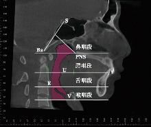
Fig.1
Upper airway boundaries"
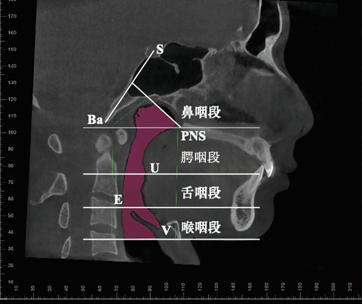
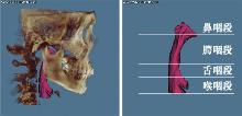
Fig.2
3D reconstruction of upper airway"
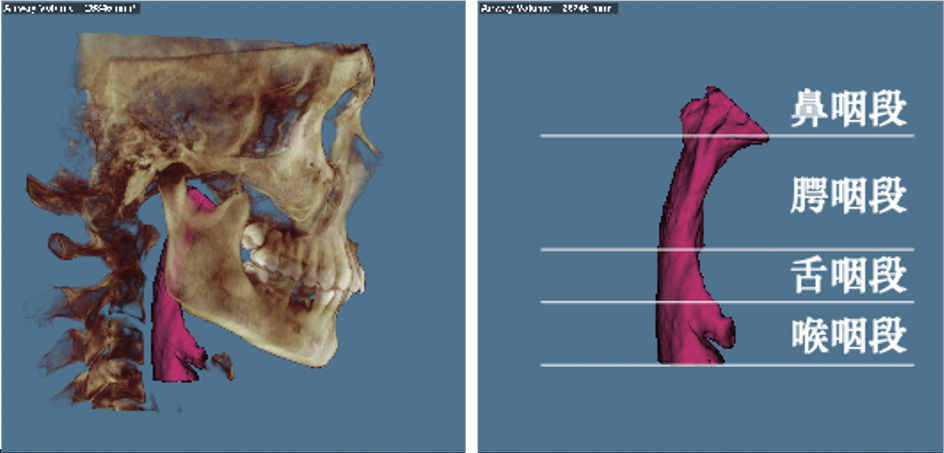
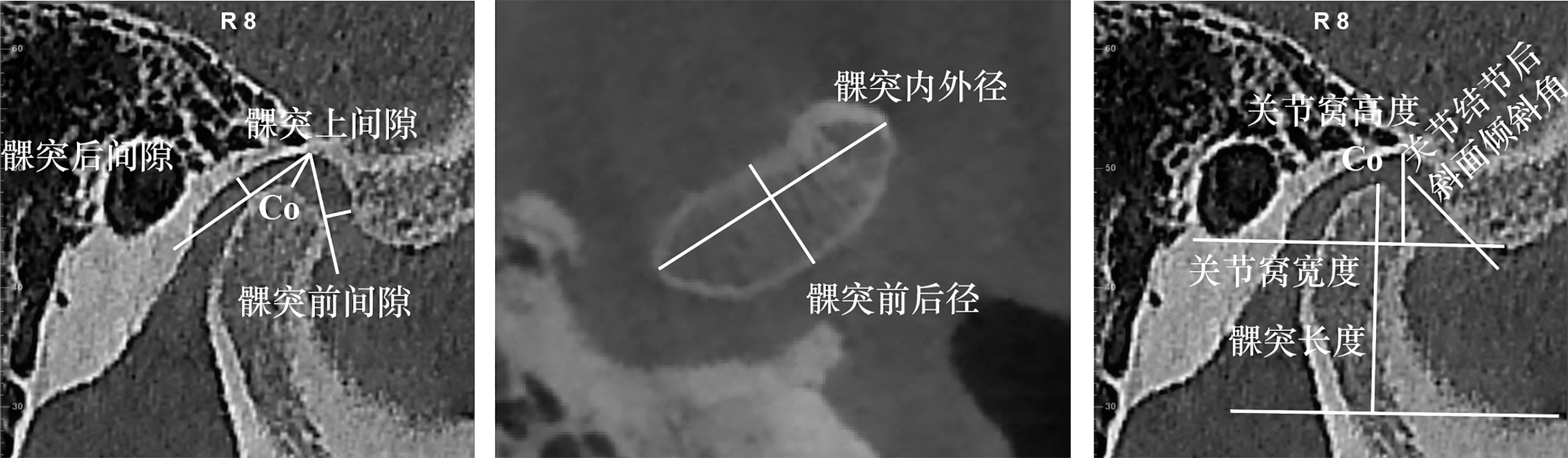
Fig.3
TMJ measurement"
Tab.1
Comparison of three-dimensional upper airway measurements in different vertical facial patterns"
| 测量项目 | 骨性Ⅱ类低角 | 骨性Ⅱ类均角 | 骨性Ⅱ类高角 | F | P |
|---|---|---|---|---|---|
| 鼻咽段体积/mm3 | 7 285.73±888.20 | 5 771.64±1 303.65 | 4 391.36±1 020.68 | 19.59 | 0.000* |
| 腭咽段体积/mm3 | 6 651.55±1 635.87 | 4 196.27±798.34 | 3 084.91±527.95 | 30.60 | 0.001* |
| 舌咽段体积/mm3 | 6 042.91±1 917.92 | 4 907.27±1 491.78 | 2 902.18±532.83 | 13.49 | 0.000* |
| 喉咽段体积/mm3 | 5 436.45±1 448.58 | 5 432.09±1 819.06 | 4 158.45±845.90 | 2.92 | 0.069 |
| 口咽最小横截面积/mm2 | 181.00±26.53 | 144.55±43.99 | 86.82±22.34 | 23.72 | 0.000* |
Tab.2
Comparison of temporomandibular joint structural characteristics in different vertical facial patterns"
| 测量项目 | 骨性Ⅱ类低角 | 骨性Ⅱ类均角 | 骨性Ⅱ类高角 | F | P | |||||
|---|---|---|---|---|---|---|---|---|---|---|
| 髁突内外径/mm | 15.74±2.12 | 18.08±2.75 | 15.79±2.45 | 3.270 | 0.051 | |||||
| 髁突前后径/mm | 7.52±0.97 | 7.32±0.83 | 6.55±0.73 | 4.022 | 0.028* | |||||
| 髁突长度/mm | 19.15±3.49 | 17.34±2.28 | 15.90±1.77 | 4.277 | 0.023* | |||||
| 关节窝宽度/mm | 15.86±1.69 | 15.21±1.58 | 16.88±1.82 | 2.691 | 0.084 | |||||
| 关节窝高度/mm | 7.35±1.52 | 6.79±1.22 | 6.76±1.12 | 0.727 | 0.492 | |||||
| 关节结节后斜面倾斜角/(°) | 52.45±11.51 | 53.67±4.42 | 53.28±8.65 | 0.057 | 0.945 | |||||
| 线性比率/% | -2.09±13.29 | -1.26±5.03 | -0.07±16.04 | 0.074 | 0.929 | |||||
Tab.3
Correlation analysis between three-dimensional upper airway measurements and temporomandibular joint structural characteristics in the hypodivergent group"
| 测量项目 | 髁突内外径 | 髁突前后径 | 髁突长度 | 关节窝宽度 | 关节窝高度 | 关节结节后 斜面倾斜角 | 线性比率 |
|---|---|---|---|---|---|---|---|
| 鼻咽体积 | -0.03 | 0.15 | 0.53 | -0.29 | -0.48 | -0.12 | -0.75* |
| 腭咽体积 | -0.24 | 0.12 | -0.19 | -0.29 | 0.24 | 0.33 | 0.00 |
| 舌咽体积 | 0.67* | 0.39 | 0.21 | 0.04 | 0.10 | 0.32 | -0.09 |
| 喉咽体积 | 0.46 | 0.22 | 0.14 | -0.30 | -0.25 | 0.33 | 0.06 |
| 口咽最小横截面积 | 0.48 | 0.32 | -0.04 | -0.15 | 0.03 | 0.53 | -0.13 |
Tab.4
Correlation analysis between three-dimensional upper airway measurements and temporomandibular joint structural characteristics in the normodivergent group"
| 测量项目 | 髁突内外径 | 髁突前后径 | 髁突长度 | 关节窝宽度 | 关节窝高度 | 关节结节后 斜面倾斜角 | 线性比率 |
|---|---|---|---|---|---|---|---|
| 鼻咽体积 | -0.31 | -0.19 | 0.16 | 0.23 | 0.01 | 0.03 | 0.24 |
| 腭咽体积 | -0.48 | -0.38 | -0.21 | -0.33 | -0.16 | -0.12 | 0.11 |
| 舌咽体积 | 0.22 | -0.13 | -0.20 | -0.18 | -0.41 | -0.06 | -0.32 |
| 喉咽体积 | 0.40 | 0.13 | -0.10 | 0.08 | -0.04 | 0.16 | -0.63* |
| 口咽最小横截面积 | -0.20 | -0.49 | -0.54 | -0.62* | -0.61* | -0.04 | 0.09 |
Tab.5
Correlation analysis between three-dimensional upper airway measurements and temporomandibular joint structural characteristics in the hyperdivergent group"
| 测量项目 | 髁突内外径 | 髁突前后径 | 髁突长度 | 关节窝宽度 | 关节窝高度 | 关节结节后 斜面倾斜角 | 线性比率 |
|---|---|---|---|---|---|---|---|
| 鼻咽体积 | 0.40 | 0.48 | 0.28 | -0.19 | 0.02 | 0.17 | -0.31 |
| 腭咽体积 | -0.20 | 0.28 | 0.08 | 0.16 | -0.06 | 0.01 | -0.44 |
| 舌咽体积 | 0.21 | -0.09 | -0.19 | 0.01 | 0.15 | 0.27 | -0.75* |
| 喉咽体积 | 0.37 | -0.07 | -0.31 | -0.29 | -0.01 | -0.07 | -0.33 |
| 口咽最小横截面积 | -0.15 | 0.26 | 0.21 | -0.12 | -0.50 | 0.37 | -0.43 |
| [1] |
Hu HN, Lin WR, Wu J, et al. Research on the correlation between the size of condyle and occlusion plane in skeletal Class Ⅱ malocclusions[J]. Clin Exp Dent Res, 2022, 8(6): 1547-1554.
doi: 10.1002/cre2.v8.6 |
| [2] |
Maffei C, de Mello MM, de Biase NG, et al. Videofluoroscopic evaluation of mastication and swallowing in individuals with TMD[J]. Braz J Otorhinolaryngol, 2012, 78(4): 24-28.
doi: S1808-86942012000400006 pmid: 22936132 |
| [3] | Shokri A, Miresmaeili A, Ahmadi A, et al. Comparison of pharyngeal airway volume in different skeletal facial patterns using cone beam computed tomography[J]. J Clin Exp Dent, 2018, 10(10):e1017. |
| [4] |
Wang TH, Yang ZH, Yang F, et al. A three dimensional study of upper airway in adult skeletal Class Ⅱ patients with different vertical growth patterns[J]. PLoS One, 2014, 9(4): e95544.
doi: 10.1371/journal.pone.0095544 |
| [5] |
Zhang Y, Che B, Ni YY, et al. Three-dimensional condylar positions and forms associated with different anteroposterior skeletal patterns and facial asymmetry in Chinese adolescents[J]. Acta Odontol Scand, 2013, 71(5): 1174-1180.
doi: 10.3109/00016357.2012.757359 pmid: 23294119 |
| [6] |
Chen YX, Li LF, Li Y, et al. Comprehensive positional and morphological assessments of the temporomandibular joint in adolescents with skeletal Class Ⅲ malocclusion: A retrospective CBCT study[J]. BMC Oral Health, 2023, 23(1): 78.
doi: 10.1186/s12903-023-02788-4 |
| [7] |
Xu JT, Sun RN, Wang LN, et al. Cone-beam evaluation of pharyngeal airway space in adult skeletal Class Ⅱ patients with different condylar positions[J]. Angle Orthod, 2019, 89(2): 312-316.
doi: 10.2319/040518-253.1 |
| [8] |
Yuen H, Rossouw PE, Wolford LM, et al. Pharyngeal airway space changes after condylar replacement and mandibular advancement surgery[J]. J Oral Maxillofac Surg, 2018, 76(6): 1165-1174.
doi: 10.1016/j.joms.2017.12.022 |
| [9] |
Nasih M, Mbyayingabo D, Li YT, et al. Exploring the relationship between condylar position and pharyngeal airway dimensions using 3D analysis across different skeletal patterns[J]. Sci Rep, 2025, 15(1): 42153.
doi: 10.1038/s41598-025-26127-5 |
| [10] | 刘扬. 成人上气道形态特征与颞下颌关节解剖结构的相关性研究[D]. 大连: 大连医科大学, 2020. |
| [11] |
Stringert HG, Worms FW. Variations in skeletal and dental patterns in patients with structural and functional alterations of the temporomandibular joint: A preliminary report[J]. Am J Orthod, 1986, 89(4): 285-297.
pmid: 3457527 |
| [12] |
Saccucci M, Polimeni A, Festa F, et al. Do skeletal cephalometric characteristics correlate with condylar volume, surface and shape? A 3D analysis[J]. Head Face Med, 2012, 8: 15.
doi: 10.1186/1746-160X-8-15 pmid: 22587445 |
| [13] |
Park IY, Kim JH, Park YH. Three-dimensional cone-beam computed tomography based comparison of condylar position and morphology according to the vertical skeletal pattern[J]. Korean J Orthod, 2015, 45(2): 66-73.
doi: 10.4041/kjod.2015.45.2.66 |
| [14] |
Santander P, Quast A, Olbrisch C, et al. Comprehensive 3D analysis of condylar morphology in adults with different skeletal patterns-A cross-sectional study[J]. Head Face Med, 2020, 16(1): 33.
doi: 10.1186/s13005-020-00245-z pmid: 33256789 |
| [15] | Yilihamu S, Li Y, Nueraihemaiti Z, et al. Comparison study of chewing efficiency in patients with different vertical skeletal patterns of Angle Class I and Angle Class Ⅱ malocclusions[J]. Quintessence Int, 2024, 55(3): 224-230. |
| [16] |
Ali Sirsmith John Z, Shrivastav SS, Kamble R, et al. Three-dimensional comparative evaluation of articular disc position and other temporomandibular joint morphology in Class Ⅱ horizontal and vertical cases with Class I malocclusion[J]. Angle Orthod, 2020, 90(5): 707-714.
doi: 10.2319/121519-801.1 pmid: 33378480 |
| [17] |
Dalili Z, Khaki N, Kia SJ, et al. Assessing joint space and condylar position in the people with normal function of temporomandib-ular joint with cone-beam computed tomography[J]. Dent Res J, 2012, 9(5): 607-612.
doi: 10.4103/1735-3327.104881 |
| [18] |
Truong L, Reher P, Doan N. Correlation between upper airway dimension and TMJ position in patients with sleep disordered breathing[J]. Cranio, 2023, 41(4): 331-339.
doi: 10.1080/08869634.2020.1853465 |
| [19] |
Ciavarella D, Ferrara D, Spinoso G, et al. Airway analysis and morphometric assessment of dental arches in obstructive sleep apnea patients[J]. J Clin Med, 2025, 14(2): 296.
doi: 10.3390/jcm14020296 |
| Viewed | ||||||
|
Full text |
|
|||||
|
Abstract |
|
|||||
