| [1] |
陈旭, 李璐, 王晓茜, 等. Ⅲ/Ⅳ期牙周炎的临床诊治决策进展[J]. 口腔医学, 2025, 45(1):13-17.
|
| [2] |
Ye ZY, Ye H, Yu XX, et al. Timing selection for loosened tooth fixation based on degree of alveolar bone resorption: A finite element analysis[J]. BMC Oral Health, 2022, 22(1): 328.
doi: 10.1186/s12903-022-02375-z
|
| [3] |
Golob Deeb J, Carrico CK, Miller A, et al. Maintenance of periodontally compromised teeth using periodontal splints[J]. Int J Dent, 2025, 2025: 7119673.
doi: 10.1155/ijod.v2025.1
|
| [4] |
周燕平, 覃沅华, 梁剑梅, 等. 高强纤维牙周夹板固定法对重度牙周炎松动牙的应用研究进展[J]. 中国医药科学, 2022, 12(18): 44-47.
|
| [5] |
Ge YC, Zhao T, Fan SZ, et al. Effects of surface treatments on the adhesion strengths between polyether ether ketone and both composite resins and poly(methyl methacrylate)[J]. BMC Oral Health, 2025, 25(1): 940.
doi: 10.1186/s12903-025-06305-7
|
| [6] |
Zhang YH, Zhang WF, Yang MY, et al. Comprehensive review of polyetheretherketone use in dentistry[J]. J Prosthodont Res, 2025, 69(2): 215-232.
doi: 10.2186/jpr.JPR_D_24_00142
pmid: 39756889
|
| [7] |
谷德奥, 陆伟. 聚芳醚酮的口腔临床应用及其产品化研究进展[J]. 中华口腔医学研究杂志(电子版), 2020, 14(4): 265-270.
|
| [8] |
Rauch A, Hahnel S, Günther E, et al. Tooth-colored CAD/CAM materials for application in 3-unit fixed dental prostheses in the molar area: An illustrated clinical comparison[J]. Materials, 2020, 13(24): 5588.
doi: 10.3390/ma13245588
|
| [9] |
葛勇城, 赵婷, 殷皓宇, 等. 聚醚醚酮在口腔医学中的数字化应用[J]. 现代口腔医学杂志, 2025, 39(2): 135-139.
|
| [10] |
冀堃, 朱顶贵, 陆伟, 等. 聚醚醚酮/聚醚酮酮材料及数字化技术在儿童口腔医学应用的初步临床报告[J]. 实用口腔医学杂志, 2020, 36(2): 402-408.
|
| [11] |
Soares Machado P, Cadore Rodrigues AC, Chaves ET, et al. Surface treatments and adhesives used to increase the bond strength between polyetheretherketone and resin-based dental materials: A scoping review[J]. J Adhes Dent, 2022, 24: b2288283.
|
| [12] |
叶颖, 邸萍, 贾胜男, 等. 个别正常牙合牙尖交错位的最大牙合力绝对值及其牙位分布的横断面研究[J]. 中华口腔医学杂志, 2015, 50(9): 536-539.
|
| [13] |
朱春晖, 虎小毅, 孙俊毅, 等. 3D打印牙周导板辅助松牙固定的临床疗效评价[J]. 临床口腔医学杂志, 2020, 36(12): 731-734.
|
| [14] |
刘延周, 李海燕, 王胧英, 等. 纤维树脂夹板固定对牙周炎伴牙齿松动患者牙周指数的影响[J]. 医师在线, 2025, 15(1): 51-53.
|
| [15] |
李双林. 树脂夹板与高强纤维牙周夹板治疗重度牙周炎伴牙齿松动患者的效果分析[J]. 当代医学, 2022, 28(2): 182-184.
|
| [16] |
Zhang CR, Liu Q, Yang JW, et al. A digital technique for splinting periodontally compromised mobile teeth in the mandibular anterior region[J]. J Prosthet Dent, 2021, 125(4): 560-563.
doi: 10.1016/j.prosdent.2020.03.004
pmid: 32414586
|
| [17] |
Wang ZY, Guo LH, Deng K, et al. Additively manufactured zirconia periodontal splint integrated prosthesis: A proof-of-concept[J]. J Dent, 2025, 156: 105701.
doi: 10.1016/j.jdent.2025.105701
|
| [18] |
Hahnel S, Wieser A, Lang R, et al. Biofilm formation on the surface of modern implant abutment materials[J]. Clin Oral Implants Res, 2015, 26(11): 1297-1301.
doi: 10.1111/clr.2015.26.issue-11
|
| [19] |
王睿瑾, 张嘉琪, 衣颖杰, 等. 牙科可切削聚醚醚酮表面抛光性能的初步研究[J]. 中华口腔医学研究杂志(电子版), 2021, 15(5): 278-283.
|
| [20] |
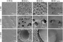
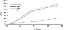
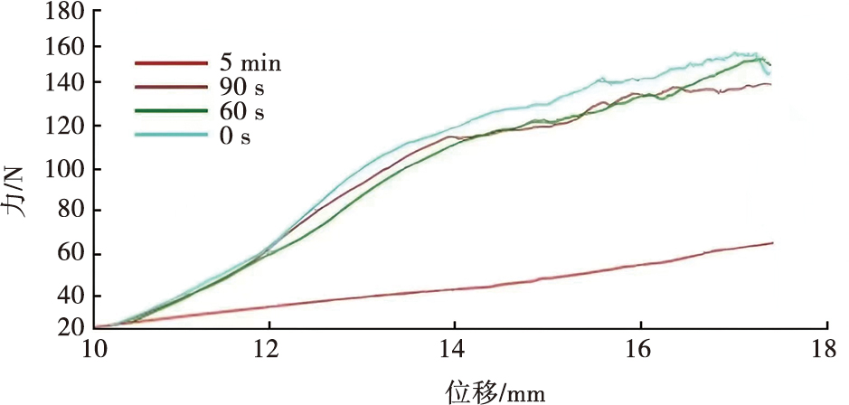
Zhang JQ, Yi YJ, Wang CW, et al. Effect of acid-etching duration on the adhesive performance of printed polyetheretherketone to veneering resin[J]. Polymers, 2021, 13(20): 3509.
doi: 10.3390/polym13203509
|
| [21] |
Sproesser O, Schmidlin PR, Uhrenbacher J, et al. Effect of sulfuric acid etching of polyetheretherketone on the shear bond strength to resin cements[J]. J Adhes Dent, 2014, 16(5): 465-472.
doi: 10.3290/j.jad.a32806
pmid: 25264546
|
| [22] |
Liu YC, Bai SZ, Zhong S, et al. Digital workflow for periodontal splinting with a guided device[J]. J Esthet Restor Dent, 2023, 35(4): 621-624.
doi: 10.1111/jerd.13026
pmid: 36810946
|
| [23] |
邵金龙, 于洋, 吕春旭, 等. 欧洲牙周病学会牙周炎治疗S3级临床指南的介绍与应用解读[J]. 中华口腔医学杂志, 2022, 57(12): 1202-1208.
|

 )
)
 苏公网安备32010602011670号