| [1] |
Huang XF, Xie MR, Xie YL, et al. The roles of osteocytes in alveolar bone destruction in periodontitis[J]. J Transl Med, 2020, 18(1): 479.
doi: 10.1186/s12967-020-02664-7
pmid: 33308247
|
| [2] |
Huja SS, Fernandez SA, Hill KJ, et al. Remodeling dynamics in the alveolar process in skeletally mature dogs[J]. Anat Rec A Discov Mol Cell Evol Biol, 2006, 288(12): 1243-1249.
|
| [3] |
Reznikov N, Shahar R, Weiner S. Bone hierarchical structure in three dimensions[J]. Acta Biomater, 2014, 10(9): 3815-3826.
doi: 10.1016/j.actbio.2014.05.024
pmid: 24914825
|
| [4] |
Nalla RK, Kinney JH, Ritchie RO. Mechanistic fracture criteria for the failure of human cortical bone[J]. Nat Mater, 2003, 2(3): 164-168.
doi: 10.1038/nmat832
pmid: 12612673
|
| [5] |
Raghavan M, Sahar ND, Kohn DH, et al. Age-specific profiles of tissue-level composition and mechanical properties in murine cortical bone[J]. Bone, 2012, 50(4): 942-953.
doi: 10.1016/j.bone.2011.12.026
pmid: 22285889
|
| [6] |
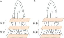
Sanz M, Marco Del Castillo A, Jepsen S, et al. Periodontitis and cardiovascular diseases: Consensus report[J]. J Clin Periodontol, 2020, 47(3): 268-288.
doi: 10.1111/jcpe.13189
pmid: 32011025
|
| [7] |
Jacobs R, Fontenele RC, Lahoud P, et al. Radiographic diagnosis of periodontal diseases: Current evidence versus innovations[J]. Periodontol 2000, 2024, 95(1): 51-69.
doi: 10.1111/prd.v95.1
|
| [8] |
Taylor EA, Donnelly E. Raman and Fourier transform infrared imaging for characterization of bone material properties[J]. Bone, 2020, 139: 115490.
doi: 10.1016/j.bone.2020.115490
|
| [9] |
Ember KJI, Hoeve MA, McAughtrie SL, et al. Raman spectroscopy and regenerative medicine: A review[J]. NPJ Regen Med, 2017, 2: 12.
doi: 10.1038/s41536-017-0014-3
pmid: 29302348
|
| [10] |
Khalid M, Bora T, Al Ghaithi A, et al. Raman spectroscopy detects changes in bone mineral quality and collagen cross-linkage in Staphylococcus infected human bone[J]. Sci Rep, 2018, 8(1): 9417.
doi: 10.1038/s41598-018-27752-z
|
| [11] |
Prats-Mateu B, Gierlinger N. Tip in-light on:Advantages, challenges, and applications of combining AFM and Raman microscopy on biological samples[J]. Microsc Res Tech, 2017, 80(1):30-40.
doi: 10.1002/jemt.v80.1
|
| [12] |
Britton M, Monahan GE, Murphy CG, et al. An investigation of composition, morphology, mechanical properties, and microdam-age accumulation of human type 2 diabetic bone[J]. Bone, 2024, 187: 117190.
doi: 10.1016/j.bone.2024.117190
|
| [13] |
Watanabe K, Lewis S, Guo XH, et al. Regional variations of jaw bone characteristics in an ovariectomized rat model[J]. J Mech Behav Biomed Mater, 2020, 110: 103952.
doi: 10.1016/j.jmbbm.2020.103952
|
| [14] |
Rubin MR, Paschalis EP, Poundarik A, et al. Advanced glycation endproducts and bone material properties in type 1 diabetic mice[J]. PLoS One, 2016, 11(5):e0154700.
|
| [15] |
Unal M, Ahmed R, Mahadevan-Jansen A, et al. Compositional assessment of bone by Raman spectroscopy[J]. Analyst, 2021, 146(24): 7464-7490.
doi: 10.1039/d1an01560e
pmid: 34786574
|
| [16] |
Khan AF, Awais M, Khan AS, et al. Raman spectroscopy of natural bone and synthetic apatites[J]. Appl Spectrosc Rev, 2013, 48(4): 329-355.
doi: 10.1080/05704928.2012.721107
|
| [17] |
Movasaghi Z, Rehman S, Rehman IU. Raman spectroscopy of biological tissues[J]. Appl Spectrosc Rev, 2007, 42(5): 493-541.
doi: 10.1080/05704920701551530
|
| [18] |
Talari ACS, Movasaghi Z, Rehman S, et al. Raman spectroscopy of biological tissues[J]. Appl Spectrosc Rev, 2015, 50(1):46-111.
doi: 10.1080/05704928.2014.923902
|
| [19] |
Unal M, Jung H, Akkus O. Novel Raman spectroscopic biomarkers indicate that postyield damage denatures bone’s collagen[J]. J Bone Miner Res, 2016, 31(5): 1015-1025.
doi: 10.1002/jbmr.2768
|
| [20] |
St Dollente Mesias V, Zhang JN, Fu WH, et al. Enhanced characterization of protein secondary structure transitions using Raman and SERS measurements combined with 2D correlation spectroscopy and principal component analysis[J]. Spectrochim Acta A Mol Biomol Spectrosc, 2025, 343: 126607.
doi: 10.1016/j.saa.2025.126607
|
| [21] |
魏洁雅, 徐思群, 周学东, 等. 牙槽骨修复重建分子调控机制的研究新进展[J]. 四川大学学报(医学版), 2024, 55(1): 31-38.
|
| [22] |
Zhou M, Graves DT. Impact of the host response and osteoblast lineage cells on periodontal disease[J]. Front Immunol, 2022, 13: 998244.
doi: 10.3389/fimmu.2022.998244
|
| [23] |
Terkawi MA, Matsumae G, Shimizu T, et al. Interplay between inflammation and pathological bone resorption: Insights into recent mechanisms and pathways in related diseases for future perspectives[J]. Int J Mol Sci, 2022, 23(3): 1786.
doi: 10.3390/ijms23031786
|
| [24] |
唐苗宁, 吴斌, 刘懋, 等. 基于单轴压缩实验初探人牙槽骨不同部位松质骨力学性能[J]. 口腔医学, 2023, 43(5): 421-426.
|
| [25] |
Wu B, Yuan L, Liu M, et al. Construction of a viscoelastic model of human cancellous bone in alveolar bone based on bone mineral density distribution[J]. Materials, 2023, 16(23): 7427.
doi: 10.3390/ma16237427
|
| [26] |
Wang FX, Zheng LY, Theopold J, et al. Methods for bone quality assessment in human bone tissue: A systematic review[J]. J Orthop Surg Res, 2022, 17(1): 174.
doi: 10.1186/s13018-022-03041-4
pmid: 35313901
|
| [27] |
Shah FA. Towards refining Raman spectroscopy-based assessment of bone composition[J]. Sci Rep, 2020, 10(1): 16662.
doi: 10.1038/s41598-020-73559-2
pmid: 33028904
|
| [28] |
Barth A. Infrared spectroscopy of proteins[J]. Biochim Biophys Acta, 2007, 1767(9): 1073-1101.
doi: 10.1016/j.bbabio.2007.06.004
pmid: 17692815
|

 ), 严斌1,2,3(
 苏公网安备32010602011670号