
Stomatology ›› 2023, Vol. 43 ›› Issue (5): 415-420.doi: 10.13591/j.cnki.kqyx.2023.05.005
• Basic Research • Previous Articles Next Articles
GE Xiao1,YU Miao2,WU Wei3,BI Xiuting4,WU Xiaoyan1,YU Chen1,LI Ti4( )
)
Revised:2023-01-18
Online:2023-05-28
Published:2023-05-31
Contact:
LI Ti
E-mail:15906368098@163.com
CLC Number:
GE Xiao,YU Miao,WU Wei,BI Xiuting,WU Xiaoyan,YU Chen,LI Ti. The effect of tantalum coating on the proliferation and osteogenic differentiation of human periodontal ligament stem cells[J]. Stomatology, 2023, 43(5): 415-420.
Tab.1
Primer sequences for qPCR"
| 基因名称 | 上游引物(5'—3') | 下游引物(5'—3') |
|---|---|---|
| ALP | GGCGGTGAACGAGAGAATGT | GGACGTAGTTCTGCTCGTGG |
| OCN | TCACACTCCTCGCCCTATTG | CTCTTCACTACCTCGCTGCC |
| RUNX2 | GGAGTGGACGAGGCAAGAGT | AGGCGGTCAGAGAACAAACT |
| GADPH | GCACCGTCAAGGCTGAGAAC | TGGTGAAGACGCCAGTGGA |
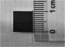
Fig.1
Appearance of Ta-coated specimen"

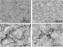
Fig.2
SEM images of the surface of SLA and Ta group specimens"

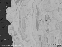
Fig.3
SEM images of Ta group specimen sections( ×2 000)"

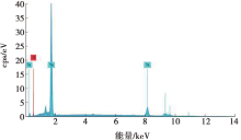
Fig.4
EDS analysis spectra of surface element of Ta group specimens"
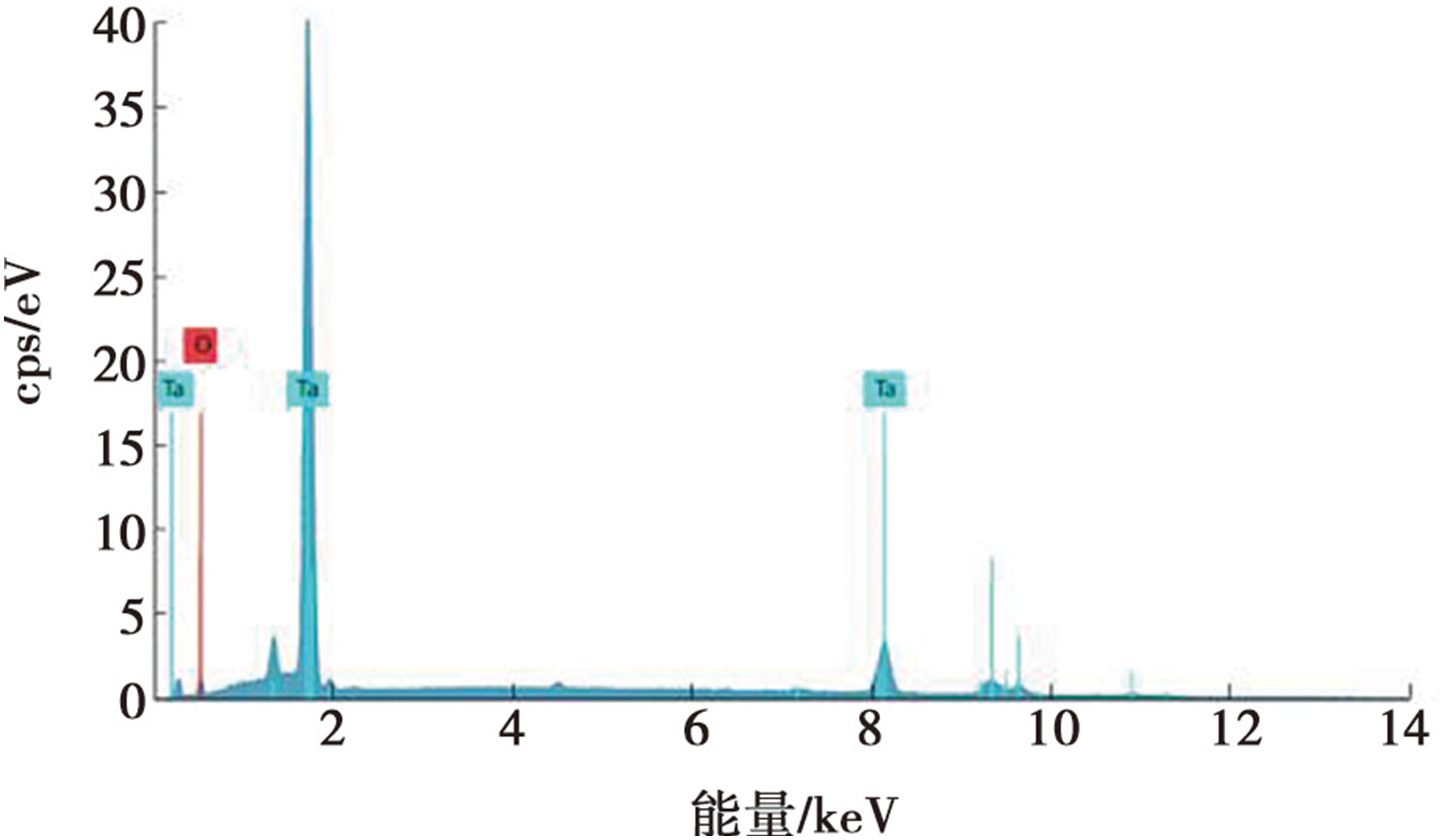
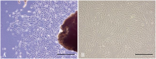
Fig.5
Microscopic morphology of hPDLSCs"

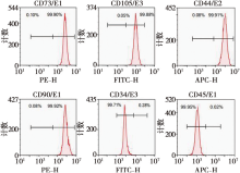
Fig.6
Surface antigens of hPDLSCs were detected by flowcytometry"

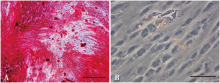
Fig.7
Osteogeogenesis and adipogenesis of hPDLSCs"

Tab.2
CCK-8 detection of hPDLSCs proliferation"
| 分组 | 1 d | 3 d | 5 d | 7 d |
|---|---|---|---|---|
| P组 | 0.15±0.01 | 0.25±0.07 | 0.35±0.06 | 1.02±0.04 |
| SLA组 | 0.15±0.00 | 0.27±0.06 | 0.40±0.03# | 1.28±0.08# |
| Ta组 | 0.16±0.00 | 0.39±0.01*# | 0.64±0.05*# | 1.70±0.21*# |
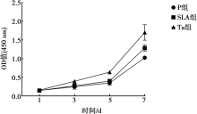
Fig.8
CCK-8 detection of hPDLSCs proliferation"
Tab.3
Alkaline phosphatase activity assay of hPDLSCs U/gprog"
| 分组 | 7 d | 14 d |
|---|---|---|
| P组 | 7.80±2.71 | 51.58±0.72 |
| SLA组 | 9.90±5.43 | 53.87±7.59 |
| Ta组 | 15.08±6.19 | 73.58±5.20**## |
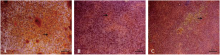
Fig.9
Alizarin red stain( ×20)"

Tab.4
Relative mRNA expression of ALP, RUNX2 and OCN"
| 组别 | ALP | RUNX2 | OCN |
|---|---|---|---|
| P组 | 1.00±0.24 | 1.00±0.09 | 1.00±0.15 |
| SLA组 | 1.47±0.17* | 1.30±0.10* | 1.49±0.10** |
| Ta组 | 1.86±0.11** | 2.44±0.30**## | 1.88±0.16**## |
| [1] |
Li HL, Yao ZG, Zhang J, et al. The progress on physicochemical properties and biocompatibility of tantalum-based metal bone implants[J]. SN Appl Sci, 2020, 2(4):671.
doi: 10.1007/s42452-020-2480-2 |
| [2] |
Mani G, Porter D, Grove K, et al. A comprehensive review of biological and materials properties of Tantalum and its alloys[J]. J Biomed Mater Res A, 2022, 110(6):1291-1306.
doi: 10.1002/jbm.a.v110.6 |
| [3] | 石晓岫, 毛世龙, 刘洋, 等. 钽与钛(合金)骨科材料的差异比较:理化指标及抗菌和成骨能力[J]. 中国组织工程研究, 2021, 25(4):593-599. |
| [4] |
Wang X, Liu WT, Yu XD, et al. Advances in surface modification of tantalum and porous tantalum for rapid osseointegration:A thematic review[J]. Front Bioeng Biotechnol, 2022, 10:983695.
doi: 10.3389/fbioe.2022.983695 |
| [5] |
吴犇, 陈晓艺, 路怀峰, 等. 等离子喷涂技术在骨植入体表面改性中的研究进展[J]. 中国表面工程, 2021, 34(4):1-11.
doi: 10.11933/j.issn.1007-9289.20210410001 |
| [6] |
Zhou X, Hu XL, Lin Y. Coating of sandblasted and acid-etched dental implants with tantalum using vacuum plasma spraying[J]. Implant Dent, 2018, 27(2):202-208.
doi: 10.1097/ID.0000000000000727 |
| [7] |
Zhu Y, Gu YX, Qiao SC, et al. Bacterial and mammalian cells adhesion to tantalum-decorated micro-/ nano-structured titanium[J]. J Biomed Mater Res A, 2017, 105(3):871-878.
doi: 10.1002/jbm.a.35953 pmid: 27784134 |
| [8] |
张成, 耿藤瑜, 王晶, 等. 纳米形貌钛种植体对骨质疏松大鼠骨结合的影响[J]. 口腔医学研究, 2022, 38(6):545-552.
doi: 10.13701/j.cnki.kqyxyj.2022.06.012 |
| [9] |
Sayed ME, Mugri MH, Almasri MA, et al. Role of stem cells in augmenting dental implant osseointegration:A systematic review[J]. Coatings, 2021, 11(9):1035.
doi: 10.3390/coatings11091035 |
| [10] |
DohanEhrenfest DM, Coelho PG, Kang BS, et al. Classification of osseointegrated implant surfaces:Materials, chemistry and topography[J]. Trends Biotechnol, 2010, 28(4):198-206.
doi: 10.1016/j.tibtech.2009.12.003 pmid: 20116873 |
| [11] |
Ding XL, Xu SL, Li SB, et al. Biological effects of titanium surface charge with a focus on protein adsorption[J]. ACS Omega, 2020, 5(40):25617-25624.
doi: 10.1021/acsomega.0c02518 pmid: 33073087 |
| [12] |
Dehghanghadikolaei A, Fotovvati B. Coating techniques for functional enhancement of metal implants for bone replacement:A review[J]. Materials (Basel), 2019, 12(11):1795.
doi: 10.3390/ma12111795 |
| [13] |
Barrak FN, Li SW, Muntane AM, et al. Particle release from implantoplasty of dental implants and impact on cells[J]. Int J Implant Dent, 2020, 6(1):50.
doi: 10.1186/s40729-020-00247-1 pmid: 32918144 |
| [14] |
Wang X, Ning BY, Pei XB. Tantalum and its derivatives in orthopedic and dental implants:Osteogenesis and antibacterial properties[J]. Colloids Surf B Biointerfaces, 2021, 208:112055.
doi: 10.1016/j.colsurfb.2021.112055 |
| [15] |
Hao JZ, Li Y, Li BE, et al. Biological and mechanical effects of micro-nanostructured titanium surface on an osteoblasticcell line in vitro and osteointegration in vivo[J]. Appl Biochem Biotechnol, 2017, 183(1):280-292.
doi: 10.1007/s12010-017-2444-1 |
| [16] |
Winning L, El Karim IA, Lundy FT. A comparative analysis of the osteogenicpotential of dental mesenchymalstem cells[J]. Stem Cells Dev, 2019, 28(15):1050-1058.
doi: 10.1089/scd.2019.0023 pmid: 31169063 |
| [17] |
Skoog SA, Kumar G, Goering PL, et al. Biological response of human bone marrow-derived mesenchymalstem cells to commercial tantalum coatings with microscale and nanoscalesurface topographies[J]. JOM, 2016, 68(6):1672-1678.
doi: 10.1007/s11837-016-1934-x |
| [18] | 蔡洪桢. 钛基人工种植体表面钽涂层改性对其生物学性能影响的实验研究[D]. 北京: 中国人民解放军医学院, 2017. |
| [19] |
Lewallen EA, Trousdale WH, Thaler R, et al. Surface roughness of titanium orthopedic implants alters the biological phenotype of human mesenchymalstromal cells[J]. Tissue Eng Part A, 2021, 27(23/24):1503-1516.
doi: 10.1089/ten.tea.2020.0369 |
| [20] |
Ma JW, Zan R, Chen WZ, et al. Cell behaviors on surface of pure tantalum with nano-dimpled structure[J]. Rare Met, 2019, 38(6):543-551.
doi: 10.1007/s12598-019-01226-1 |
| [21] |
Marconi GD, Fonticoli L, Della Rocca Y, et al. Human periodontal ligament stem cells response to titanium implant surface:Extracellular matrix deposition[J]. Biology, 2021, 10(9):931.
doi: 10.3390/biology10090931 |
| [22] |
Shi JY, Zhang XM, Qiao SC, et al. Enhanced osteointegration of tantalum-modified titanium implants with micro/nano-topography[J]. RSC Adv, 2017, 7(73):46472-46479.
doi: 10.1039/C7RA08036K |
| [23] |
An R, Fan PP, Zhou MJ, et al. Nanolamellartantalum interfaces in the osteoblast adhesion[J]. Langmuir, 2019, 35(7):2480-2489.
doi: 10.1021/acs.langmuir.8b02796 |
| [24] | 姜铭坤, 何亚茹, 吴文慧. 钛钽生物梯度材料在成骨过程中对碱性磷酸酶的影响[J]. 当代医学, 2021, 27(6):39-41. |
| [25] |
Piglionico S, Bousquet J, Fatima N, et al. Porous tantalum VS. titaniumimplants:Enhanced mineralized matrix formation after stem cells proliferation and differentiation[J]. J Clin Med, 2020, 9(11):3657.
doi: 10.3390/jcm9113657 |
| [26] |
Liu XY, Song XB, Zhang P, et al. Effects of nano tantalum implants on inducing osteoblast proliferation and differentiation[J]. Exp Ther Med, 2016, 12(6):3541-3544.
doi: 10.3892/etm.2016.3801 pmid: 28101149 |
| [27] |
Vimalraj S. Alkaline phosphatase:Structure, expression and its function in bone mineralization[J]. Gene, 2020, 754:144855.
doi: 10.1016/j.gene.2020.144855 |
| [28] |
Komori T. Functions of osteocalcin in bone, pancreas, testis, and muscle[J]. Int J Mol Sci, 2020, 21(20):7513.
doi: 10.3390/ijms21207513 |
| [29] |
Komori T. Whole aspect of Runx2 functions in skeletal develop-ment[J]. Int J Mol Sci, 2022, 23(10):5776.
doi: 10.3390/ijms23105776 |
| [30] | Qian H, Lei T, Ye ZM, et al. From the performance to the essence:The biological mechanisms of how tantalum contributes to osteogenesis[J]. Biomed Res Int, 2020, 2020:5162524. |
| Viewed | ||||||
|
Full text |
|
|||||
|
Abstract |
|
|||||