
Stomatology ›› 2024, Vol. 44 ›› Issue (12): 892-898.doi: 10.13591/j.cnki.kqyx.2024.12.003
• Basic and Clinical Research • Previous Articles Next Articles
GUO Songsong1,2,3,ZHANG Zhenxing1,2,3,ZHANG Ping1,2,3,JIANG Chenghui1,2,3,CHENG Jie1,2,3,JIANG Hongbing1,2,3,LI Sheng1,2,3( )
)
Received:2024-08-22
Online:2024-12-28
Published:2024-12-26
CLC Number:
GUO Songsong, ZHANG Zhenxing, ZHANG Ping, JIANG Chenghui, CHENG Jie, JIANG Hongbing, LI Sheng. Evaluation of two surgical methods for maxillary hypoplasia in patients with cleft lip and palate[J]. Stomatology, 2024, 44(12): 892-898.
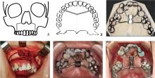
Fig.1
Schematic diagram and intraoperative photo of AMSDO surgery"


Fig.2
Anatomical landmarks of the maxilla and palatopharyngeal structure"
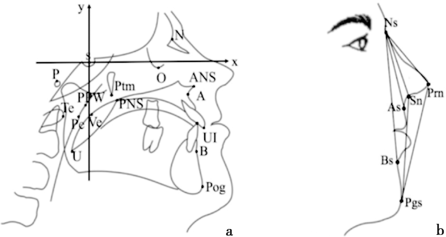
Tab.1
Composition and meaning of 18 measurement indicators"
| 类型 | 测量项目 | 含义 |
|---|---|---|
| 上颌骨改变 | Ax/mm | A点在X轴上的坐标 |
| SNA/(°) | S-N-A形成的角度 | |
| ANS-Ptm/mm | ANS与Ptm点在X轴上投影点间的距离 | |
| SNPP/(°) | ANS-PNS平面与SN平面的交角 | |
| UI-SN/(°) | 上切牙长轴与SN平面的交角 | |
| 硬组织侧貌 | SNB/(°) | S-N-B形成的角度 |
| ANB/(°) | SNA与SNB角的差值 | |
| NA-PA/(°) | NA连线与PA连线的交角 | |
| 覆盖/mm | UI点与LI点在X轴上的投影点间的差值 | |
| 软组织侧貌 | Ns-Prn-Pgs/(°) | Ns-Prn-Pgs形成的角度 |
| As-Ns-Bs/(°) | As-Ns-Bs形成的角度 | |
| Ns-Sn-Pgs/(°) | Ns-Sn-Pgs形成的角度,反映面型的凸度 | |
| 腭咽部结构 | 软腭长度/mm | PNS与U的距离 |
| 软腭厚度/mm | PNS-U的垂线与软腭最宽处两点相交形成的距离 | |
| 软硬腭交角/(°) | ANS-PNS平面与PNS-U平面的交角 | |
| 咽腔深度/mm | PNS与PPW的距离 | |
| 腭咽间隙/mm | Ve与Pe的距离 | |
| 骨性腭咽间隙/mm | Ve与Te的距离 |
Tab.2
Comparison of maxillary bone indicators one month after surgery versus one week before surgery in 25 patients with cleft lip and palate"
| 测量项目 | AMSDO(T2-T1) | Le FortⅠ型 截骨术(T2-T1) | P |
|---|---|---|---|
| Ax/mm | 6.91±1.73 | 2.82±0.69 | 0.00a |
| SNA/(°) | 7.74±1.65 | 5.01±0.98 | 0.00a |
| ANS-Ptm/mm) | 10.68±2.13 | 3.02±0.69 | 0.00a |
| SNPP/(°) | -(3.11±1.64) | 3.23±2.82 | 0.00a |
| UI-SN/(°) | 3.62±3.23 | 0.82±0.91 | 0.00a |
Tab.3
Analysis of lateral facial indicators one month after surgery versus one week before surgery in patients with cleft lip and palate"
| 测量项目 | AMSDO(T2-T1) | Le FortⅠ型 截骨术(T2-T1) | P |
|---|---|---|---|
| 覆盖/mm | 11.02±1.82 | 8.21±1.39 | 0.00a |
| SNB/(°) | -(0.41±0.65) | -(4.82±1.75) | 0.00a |
| ANB/(°) | 7.32±1.86 | 9.61±2.02 | 0.00a |
| NA-PA/(°) | 13.82±1.94 | 14.81±3.66 | 0.22 |
| Ns-Prn-Pgs/(°) | 10.35±4.85 | 7.15±2.82 | 0.02b |
| As-Ns-Bs/(°) | 6.21±2.28 | 8.89±2.06 | 0.00a |
| Ns-Sn-Pgs/(°) | 2.25±1.60 | 6.65±2.08 | 0.00a |

Fig.3
Patient in the AMSDO Group"

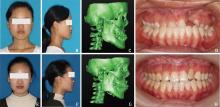
Fig.4
Patient in the LFI group"

Tab.4
Analysis of the palatopharyngeal anatomical structure in patients with cleft lip and palate, comparing 1 month post-operation to 1 week pre-operation"
| 测量项目 | AMSDO组 | Le FortⅠ组 | P |
|---|---|---|---|
| 软腭长度/mm | 0.65±0.69 | 2.01±1.71 | 0.01b |
| 咽腔深度/mm | 0.89±0.92 | 3.06±1.35 | 0.00a |
| 软硬腭交角/(°) | 5.62±2.23 | 10.23±3.11 | 0.00a |
| 软腭厚度/mm | -(0.15±0.14) | -(0.98±0.50) | 0.03b |
| 腭咽间隙/mm | 0.06±0.10 | 0.39±0.33 | 0.00a |
| 骨性腭咽间隙/mm | 0.12±0.16 | 0.58±0.62 | 0.02 b |
Tab.5
Palatopharyngeal closure and speech assessment rating scale"
| 测量项目 | AMSDO组 | Le FortⅠ组 | ||||
|---|---|---|---|---|---|---|
| 术前1周 | 术后1个月 | P | 术前1周 | 术后1个月 | P | |
| 鼻音 | 1.60±0.22 | 1.70±0.21 | 0.75 | 2.07±0.18 | 2.00±0.20 | 0.80 |
| 语音可理解性 | 1.80±0.25 | 1.70±0.26 | 0.78 | 2.47±0.19 | 2.40±0.19 | 0.80 |
| 腭咽闭合功能 | 1.90±0.18 | 1.90±0.18 | 1.00 | 1.87±0.13 | 1.80±0.14 | 0.73 |
| 呼吸习惯 | 1.80±0.13 | 1.80±0.13 | 1.00 | 1.87±0.09 | 1.87±0.09 | 1.00 |
| [1] | 戴俊峰, 邢树林. 先天性唇腭裂病因学研究的新进展[J]. 中国优生与遗传杂志, 2008, 16(12):124-125. |
| [2] | Good PM, Mulliken JB, Padwa BL. Frequency of Le Fort I osteotomy after repaired cleft lip and palate or cleft palate[J]. Cleft Palate Craniofac J, 2007, 44(4):396-401. |
| [3] |
Mossey PA, Little J, Munger RG, et al. Cleft lip and palate[J]. Lancet, 2009, 374(9703):1773-1785.
doi: 10.1016/S0140-6736(09)60695-4 pmid: 19747722 |
| [4] | Prada Madrid JR, Gómez Prada DC, Gutierrez Rodríguez EP, et al. Lefort I osteotomy with and without osteogenic distraction in cleft lip and palate patients: Experience at hospital universitario infantil de San josé[J]. J Craniofac Surg, 2024, 35(3):721-725. |
| [5] | Chang CS, Wallace CG, Hsiao YC, et al. Airway changes after cleft orthognathic surgery evaluated by three-dimensional computed tomography and overnight polysomnographic study[J]. Sci Rep, 2017, 7(1):12260. |
| [6] | Venkategowda PR, Prakash AT, Roy ET, et al. Stability of vertical, horizontal and angular parameters following superior repositioning of maxilla by le fort I osteotomy: A cephalometric study[J]. J Clin Diagn Res, 2017, 11(1):ZC10-ZC14. |
| [7] | Bengi O, Karaçay S, Akin E, et al. Cephalometric evaluation of patients treated by maxillary anterior segmental distraction: A preliminary report[J]. J Craniomaxillofac Surg, 2007, 35(6/7):302-310. |
| [8] | Gunaseelan R, Cheung LK, Krishnaswamy R, et al. Anterior maxillary distraction by tooth-borne palatal distractor[J]. J Oral Maxillofac Surg, 2007, 65(5):1044-1049. |
| [9] | Richardson S, Selvaraj D, Khandeparker RV, et al. Tooth-borne anterior maxillary distraction for cleft maxillary hypoplasia: Our experience with 147 patients[J]. J Oral Maxillofac Surg, 2016, 74(12):2504.e1-2504.e14. |
| [10] |
Jia HC, Zhuang L, Zhang N, et al. Comparison of skeletal maxillary transverse deficiency treated by microimplant-assisted rapid palatal expansion and tooth-borne expansion during the post-pubertal growth spurt stage[J]. Angle Orthod, 2021, 91(1):36-45.
doi: 10.2319/041920-332.1 pmid: 33289835 |
| [11] | 杨辛, 沈国芳, 张志愿, 等. 唇腭裂患者牵张成骨术后的侧貌变化[J]. 中国口腔颌面外科杂志, 2009, 7(2):97-101. |
| [12] | 郭秀娟, 耿海霞, 郑海英, 等. 上颌骨Le Fort Ⅰ型截骨前徙术对唇腭裂患者腭咽结构及功能影响的临床探讨[J]. 临床口腔医学杂志, 2017, 33(11):664-667. |
| [13] |
Jordan HN, Schenck GC, Ellis C, et al. Examining velopharyngeal closure patterns based on anatomic variables[J]. J Craniofac Surg, 2017, 28(1):270-274.
doi: 10.1097/SCS.0000000000003284 pmid: 27941550 |
| [14] |
Li HL, Dai JW, Si JW, et al. Anterior maxillary segmental distraction in the treatment of severe maxillary hypoplasia secondary to cleft lip and palate[J]. Int J Clin Exp Med, 2015, 8(9):16022-16028.
pmid: 26629107 |
| [15] | Fariña R, Lolas J, Moreno E, et al. Cleft lip and palate midfacial hypoplasia: Criteria to choose the treatment[J]. J Craniofac Surg, 2022, 33(2):496-501. |
| [16] | Kim YK. Complications associated with orthognathic surgery[J]. J Korean Assoc Oral Maxillofac Surg, 2017, 43(1):3-15. |
| [17] |
Jiang Y, Jiang CY, Shi B, et al. Efficacy of modified anterior maxillary segmental distraction osteogenesis based on 3D visualisation for the treatment of maxillary hypoplasia among adolescents with cleft lip and palate[J]. BMC Oral Health, 2024, 24(1):1032.
doi: 10.1186/s12903-024-04828-z pmid: 39227941 |
| [18] | Varidel A, Padwa BL, Britt MC, et al. Patient-specific le fort I osteotomy plates are more stable than stock plates in patients with cleft lip and palate[J/OL]. Plast Reconstr Surg, 2024[2024-08-22]. https://pubmed.ncbi.nlm.nih.gov/38546729/. |
| [19] | Chen Q, Li Y, Shi B, et al. Analysis of the correlative factors for velopharyngeal closure of patients with cleft palate after primary repair[J]. Oral Surg Oral Med Oral Pathol Oral Radiol, 2013, 116(6):e424-428 |
| [20] |
Kanzaki H, Imai Y, Nakajo T, et al. Midfacial changes through anterior maxillary distraction osteogenesis in patients with cleft lip and palate[J]. J Craniofac Surg, 2017, 28(4):1057-1062.
doi: 10.1097/SCS.0000000000003506 pmid: 28141644 |
| [21] | Yatabe-Ioshida MS, Campos LD, Yaedu RY, et al. Upper airway 3D changes of patients with cleft lip and palate after orthognathic surgery[J]. Cleft Palate Craniofac J, 2019, 56(3):314-320. |
| [22] |
Shi B, Losee JE. The impact of cleft lip and palate repair on maxillofacial growth[J]. Int J Oral Sci, 2015, 7(1):14-17.
doi: 10.1038/ijos.2014.59 pmid: 25394591 |
| [23] |
Jung J, Lee CH, Lee JW, et al. Three dimensional evaluation of soft tissue after orthognathic surgery[J]. Head Face Med, 2018, 14(1):21.
doi: 10.1186/s13005-018-0179-z pmid: 30290762 |
| [24] | Tanikawa C, Hirata K, Aikawa T, et al. Efficacy of maxillary anterior segmental distraction osteogenesis in patients with cleft lip and palate[J]. Cleft Palate Craniofac J, 2018, 55(10):1375-1381. |
| [25] | Gateno J, Engel ER, Teichgraeber JF, et al. A new Le Fort I internal distraction device in the treatment of severe maxillary hypoplasia[J]. J Oral Maxillofac Surg, 2005, 63(1):148-154. |
| Viewed | ||||||
|
Full text |
|
|||||
|
Abstract |
|
|||||