
Stomatology ›› 2024, Vol. 44 ›› Issue (4): 307-311.doi: 10.13591/j.cnki.kqyx.2024.04.013
• Summary • Previous Articles Next Articles
YAO Chengliang,WU Xiuping( )
)
Received:2022-11-24
Online:2024-04-28
Published:2024-04-25
CLC Number:
YAO Chengliang, WU Xiuping. Correlation between imaging measurements and diagnosis of adenoid hypertrophy and orthodontic treatment in adolescent children[J]. Stomatology, 2024, 44(4): 307-311.
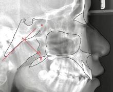
Fig.1
The fixed-point method of ZHANG Baohua"

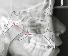
Fig.2
The fixed-point method of ZOU Mingshun"
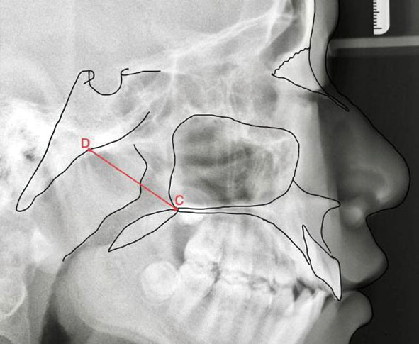
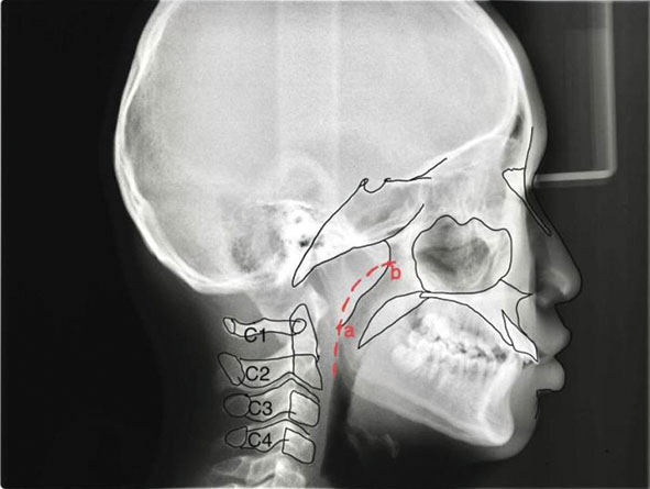
Fig.3
Parallel curve method"
| [1] | Hai W. Chronic adenoiditis[J]. J Int Med Res, 2020, 48(11):030006052097145. |
| [2] |
Orji FT, Ezeanolue BC. Evaluation of adenoidal obstruction in children: Clinical symptoms compared with roentgenographicassessment[J]. J Laryngol Otol, 2008, 122(11):1201-1205.
doi: 10.1017/S0022215108001916 pmid: 18394205 |
| [3] |
Liu JL, Li SH, Cai YM, et al. Automated radiographic evaluation of adenoid hypertrophy based on VGG-lite[J]. J Dent Res, 2021, 100(12):1337-1343.
doi: 10.1177/00220345211009474 |
| [4] |
Pereira L, Monyror J, Almeida FT, et al. Prevalence of adenoid hypertrophy: A systematic review and meta-analysis[J]. Sleep Med Rev, 2018, 38:101-112.
doi: S1087-0792(16)30137-X pmid: 29153763 |
| [5] | Iwasaki T, Hayasaki H, Takemoto Y, et al. Oropharyngeal airway in children with Class Ⅲ malocclusion evaluated by cone-beam computed tomography[J]. Am J Orthod Dentofacial Orthop, 2009, 136(3):318.e1-318.e9;discussion 318-319. |
| [6] |
Joseph AA, Elbaum J, Cisneros GJ, et al. A cephalometric comparative study of the soft tissue airway dimensions in persons with hyperdivergent and normodivergent facial patterns[J]. J Oral Maxillofac Surg, 1998, 56(2):135-139;discussion 139-140.
doi: 10.1016/S0278-2391(98)90850-3 |
| [7] |
Mattos CT, Cruz CV, da Matta TCS, et al. Reliability of upper airway linear, area, and volumetric measurements in cone-beam computed tomography[J]. Am J Orthod Dentofacial Orthop, 2014, 145(2):188-197.
doi: 10.1016/j.ajodo.2013.10.013 |
| [8] | Zhang LP, Liu H. Influence of adenoid hypertrophy on malocclusion and maxillofacial development in children[J]. Evid Based Complement Alternat Med, 2022, 2022:2052359. |
| [9] |
Solow B, Siersbaek-Nielsen S, Greve E. Airway adequacy, head posture, and craniofacial morphology[J]. Am J Orthod, 1984, 86(3):214-223.
doi: 10.1016/0002-9416(84)90373-7 pmid: 6591801 |
| [10] |
Lione R, Buongiorno M, Franchi L, et al. Evaluation of maxillary arch dimensions and palatal morphology in mouth-breathing children by using digital dental casts[J]. Int J Pediatr Otorhinolaryngol, 2014, 78(1):91-95.
doi: 10.1016/j.ijporl.2013.09.028 |
| [11] |
Festa P, Mansi N, Varricchio AM, et al. Association between upper airway obstruction and malocclusion in mouth-breathing children[J]. Acta Otorhinolaryngol Ital, 2021, 41(5):436-442.
doi: 10.14639/0392-100X-N1225 pmid: 34734579 |
| [12] | Ceccanti G, Caruso S, Pasini M, et al. Facial skeletal alterations in mouth breathing paediatric patients: Cephalometric evaluations[J]. J Biol Regul Homeost Agents, 2020, 34(1 Suppl. 1):23-32. |
| [13] | Medeiros da Fonsêca JD, Aliverti A, Benício K, et al. Breathing pattern and muscle activity using different inspiratory resistance devices in children with mouth breathing syndrome[J]. ERJ Open Res, 2022, 8(2):00480-02021. |
| [14] |
Shavakhi M, Mohamadian F, ZarifNajafi H. The effects of the headgear therapy on the airway dimensions in patients with Class Ⅱ malocclusion: A systematic review[J]. Dent Med Probl, 2019, 56(2):191-196.
doi: 10.17219/dmp/105957 |
| [15] |
Xie JY, Huang CR, Yin K, et al. Effects of orthodontic treatment with activator appliance on patients with skeletal Class II malocclusion: A systematic review and meta-analysis[J]. Ann Palliat Med, 2021, 10(12):12319-12334.
doi: 10.21037/apm-21-3205 pmid: 35016488 |
| [16] |
Arens R, McDonough JM, Corbin AM, et al. Upper airway size analysis by magnetic resonance imaging of children with obstructive sleep apnea syndrome[J]. Am J Respir Crit Care Med, 2003, 167(1):65-70.
doi: 10.1164/rccm.200206-613OC |
| [17] | 沈翎, 林宗通, 许杨杨, 等. 儿童OSAHS与腺样体和扁桃体大小的关系探讨[J]. 临床耳鼻咽喉头颈外科杂志, 2014, 28(6):381-385. |
| [18] |
Marcus CL, Brooks LJ, Draper KA, et al. Diagnosis and management of childhood obstructive sleep apnea syndrome[J]. Pediatrics, 2012, 130(3):576-584.
doi: 10.1542/peds.2012-1671 pmid: 22926173 |
| [19] |
Schechter MS. Technical report: Diagnosis and management of childhood obstructive sleep apnea syndrome[J]. Pediatrics, 2002, 109(4):e69.
doi: 10.1542/peds.109.4.e69 pmid: 11927742 |
| [20] | 曾国辉, 李郧, 滕尧树, 等. 动态腺样体-鼻咽腔比率在OSAHS患儿疾病评估中的应用研究[J]. 临床耳鼻咽喉头颈外科杂志, 2017, 31(15):1174-1177. |
| [21] |
Chung Leng Muñoz I, BeltriOrta P. Comparison of cephalometric patterns in mouth breathing and nose breathing children[J]. Int J Pediatr Otorhinolaryngol, 2014, 78(7):1167-1172.
doi: 10.1016/j.ijporl.2014.04.046 |
| [22] | Rakosi T, Schilli W. Class Ⅲ anomalies: A coordinated approach to skeletal, dental, and soft tissue problems[J]. J Oral Surg, 1981, 39(11):860-870. |
| [23] | Jha AK, Chandra S. Early management of Class Ⅲ malocclusion in mixed dentition[J]. Int J Clin Pediatr Dent, 2021, 14(2):331-334. |
| [24] |
Moideen SP, Mytheenkunju R, Govindan Nair A, et al. Role of adenoid-nasopharyngeal ratio in assessing adenoid hypertrophy[J]. Indian J Otolaryngol Head Neck Surg, 2019, 71(Suppl 1):469-473.
doi: 10.1007/s12070-018-1359-7 |
| [25] | Pagella F, Pusateri A, Chu F, et al. Adenoid assessment in paediatric patients: The role of flexible nasal endoscopy[J]. Int J ImmunopatholPharmacol, 2011, 24(S4):49-54. |
| [26] |
Önal M, YIlmaz T, Bilgiç E, et al. Possible role of apoptosis in pathogenesis of adenoid hypertrophy and chronic adenoiditis: Prospective case-control study[J]. Auris Nasus Larynx, 2015, 42(6):449-452.
doi: 10.1016/j.anl.2015.04.012 pmid: 26003878 |
| [27] | 邹明舜. 儿童增殖腺-鼻咽腔比率测定的临床价值[J]. 中华放射学杂志, 1997, 31(3):190-192. |
| [28] |
Baldassari CM, Choi S. Assessing adenoid hypertrophy in children: X-ray or nasal endoscopy?[J]. Laryngoscope, 2014, 124(7):1509-1510.
doi: 10.1002/lary.v124.7 |
| [29] |
Jana M, Gupta AK. Novel use of ultrasound in evaluation of adenoid hypertrophy in children[J]. Indian J Pediatr, 2020, 87(11):885-886.
doi: 10.1007/s12098-020-03487-6 |
| [30] | 廖昕, 陈卫国, 程勇, 等. 儿童腺样体肥大的X线诊断分析(附120例报告)[J]. 实用放射学杂志, 2007, 23(9):1239-1241. |
| [31] |
Wang YJ, Jiao HJ, Mi CR, et al. Evaluation of adenoid hypertrophy with ultrasonography[J]. Indian J Pediatr, 2020, 87(11):910-915.
doi: 10.1007/s12098-020-03203-4 |
| [32] |
Major MP, Witmans M, El-Hakim H, et al. Agreement between cone-beam computed tomography and nasoendoscopy evaluations of adenoid hypertrophy[J]. Am J Orthod Dentofacial Orthop, 2014, 146(4):451-459.
doi: 10.1016/j.ajodo.2014.06.013 |
| [33] |
Gao DK, Sun XY, Yang Y, et al. Diagnostic value of CBCT in Chinese children with adenoid hypertrophy[J]. Laryngoscope Investig Oto, 2022, 7(5):1308-1314.
doi: 10.1002/lio2.v7.5 |
| [34] |
Surov A, Ryl I, Bartel-Friedrich S, et al. MRI of nasopharyngeal adenoid hypertrophy[J]. Neuroradiol J, 2016, 29(5):408-412.
doi: 10.1177/1971400916665386 pmid: 27531860 |
| [35] | 邓莹莹, 陈观尚, 郑启文, 等. CT和MRI在检查儿童腺样体肥大(AH)中的价值比较[J]. 现代医用影像学, 2019, 28(12):2651-2653. |
| [36] |
Kurien M, Lepcha A, Mathew J, et al. X-Rays in the evaluation of adenoid hypertrophy: It’s role in the endoscopic era[J]. Indian J Otolaryngol Head Neck Surg, 2005, 57(1):45-47.
doi: 10.1007/BF02907627 |
| [37] |
Major MP, Flores-Mir C, Major PW. Assessment of lateral cephalometric diagnosis of adenoid hypertrophy and posterior upper airway obstruction: A systematic review[J]. Am J Orthod Dentofacial Orthop, 2006, 130(6):700-708.
doi: 10.1016/j.ajodo.2005.05.050 |
| [38] | 张宝华, 付孝根. DR在评价儿童腺样体肥大中的临床价值[J]. 医疗装备, 2009, 22(2):55-57. |
| [39] |
Fujioka M, Young LW, Girdany BR. Radiographic evaluation of adenoidal size in children: Adenoidal-nasopharyngeal ratio[J]. AJR Am J Roentgenol, 1979, 133(3):401-404.
doi: 10.2214/ajr.133.3.401 |
| [40] |
Yueniwati Y, Halim N. Diagnostic test value of assessment adenoid enlargement with and without airway obstruction using lateral soft tissues X-ray compared to nasoendoscopy[J]. Indian J Otolaryngol Head Neck Surg, 2019, 71(S3):1739-1744.
doi: 10.1007/s12070-017-1089-2 |
| [41] | 李东辉, 任甄华, 王晓曼, 等. 小儿腺样体肥大的X线表现(附136例总结及与132例正常对照)[J]. 临床放射学杂志, 1999, 18(11):694-697. |
| [42] | 王艳春, 林美金. 鼻咽部侧位片对腺样体肥大的诊断价值[J]. 中国误诊学杂志, 2008, 8(13):3088-3089. |
| [43] | 袁林秀. 鼻咽侧位X线片在儿童腺样体肥大诊断中的应用价值[J]. 中国实用医刊, 2013, 40(2):119-120. |
| [1] | JIN Min, CHEN Yumao, HAN Minxuan, LU Shijun. Application progress of digital indirect bonding technology in orthodontic bracket positioning [J]. Stomatology, 2024, 44(3): 227-231. |
| [2] | TIAN Xin, WU Sihua, DAN Hongxia, ZENG Xin, WANG Jiongke, CHEN Qianming. New developments in the clinical presentation and diagnosis of granulomatosis with polyangiitis [J]. Stomatology, 2024, 44(3): 209-213. |
| [3] | FU Kaiyuan, LEI Jie. Update of the classification, diagnosis and management of temporomandibular disorders [J]. Stomatology, 2024, 44(1): 6-10. |
| [4] | WANG Yong, WANG Lin. Comments on several basic concepts related to mechanics in orthodontics [J]. Stomatology, 2024, 44(1): 60-62. |
| [5] | WEI Lili, LI Bo, CHENG Yong. Advances in the clinical application of MRI in temporomandibular joint disorders [J]. Stomatology, 2024, 44(1): 11-15. |
| [6] | ZHAO Ning, FANG Bing. Research progress of orthodontics and temporomandibular disorders [J]. Stomatology, 2024, 44(1): 20-23. |
| [7] | KANG Fujia, ZHANG Xiya, YU Lei, WANG Songqing, ZHANG Haoyan, LI Xinyi, ZHU Xianchun. Three-dimensional finite element analysis of maxillary anterior teeth intrusionby clear aligner assisted with micro-implant [J]. Stomatology, 2023, 43(9): 796-802. |
| [8] | ZHOU Bin, WU Xiaoyong, LIU Mingyan. Effect of invisible functional appliance in the treatment of skeletal Class Ⅱ malocclusion in adolescents [J]. Stomatology, 2023, 43(7): 643-646. |
| [9] | XU Qingqing, WANG Yumeng, LI Mengyuan, ZHANG Yuerong, XU Hai, JIANG Fei, ZHANG Guangdong. Correlation analysis of morphological features with root canal anatomy of mandibular first premolars [J]. Stomatology, 2023, 43(7): 625-632. |
| [10] | WU Chunlan, HU Yue, WANG Tingting, YU Dedong, DENG Gang, LI Chaolun. The consistency studyon different methods of oral vestibular groove depth measurements [J]. Stomatology, 2023, 43(6): 512-517. |
| [11] | WEI Zhifeng, YANG Tongtong, WANG Jinghong. Progress of research on factors of upper airway obstruction affecting maxillofacial development [J]. Stomatology, 2023, 43(6): 573-576. |
| [12] | LI Xueying,ZOU Xiaoying,YUE Lin. Predisposing factors, clinical diagnosis and treatment strategy of tooth resorption [J]. Stomatology, 2023, 43(4): 289-293. |
| [13] | YANG Xia, YE Qingyuan, SONG Changlong, HOU Rui, YANG Yaowu, WEI Jianhua. Progress of research on diagnosis and treatment of the first bite syndrome [J]. Stomatology, 2023, 43(2): 188-192. |
| [14] | FAN Yongjie,KOU Yating. Effect of different tips of posteriors in extraction treatment on three-dimensional forces of teeth with clear aligners [J]. Stomatology, 2023, 43(2): 104-109. |
| [15] | ZHANG Tianyu,JIANG Lu. Research progress of IgG4-related disease in oral cavity [J]. Stomatology, 2023, 43(12): 1106-1111. |
| Viewed | ||||||
|
Full text |
|
|||||
|
Abstract |
|
|||||